Pharmaceutical Microbiology - Unit 3
Syllabus
Study of morphology, classification, reproduction/replication and cultivation of Fungi and Viruses. Classification and mode of action of disinfectants Factors influencing disinfection, antiseptics and their evaluation. For bacteriostatic and bactericidal actions Evaluation of bactericidal & Bacteriostatic. Sterility testing of products (solids, liquids, ophthalmic and other sterile products) according to IP, BP and USP.
Scroll to Download
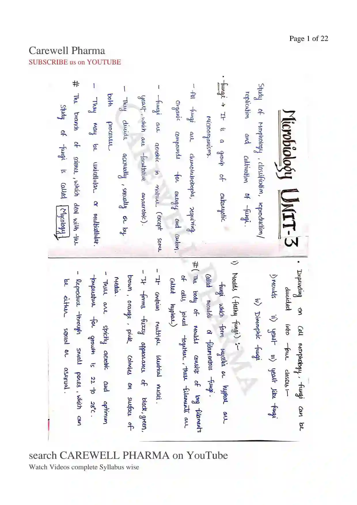
MICROBIOLOGY UNIT-3
Fungi
- It is a group of eukaryotic Microorganisms.
- All fungi are chemoheterotrophs, requiring organic compounds for energy and carbon.
- Fungi are aerobic in nature (except some yeast, which are facultative anaerobic).
- They may be unicellular or multicellular. They reproduce both asexually, sexually or by both processes.
- The branch of science, which deal with the study of fungi is called Mycology.
Depending on cell morphology, fungi can be divided into four classes
i) Moulds
ii) Yeasts
iii) Yeast Like Fungi
iv) Dimorphic Fungi
i) Moulds (Fleshy Fungi)
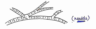
Fungi which form mycelia or hyphae are called moulds or filamentous fungi.
- (The body of moulds consists of long filaments of cells joined together, these filaments are called hyphae)
- It contain multiple identical nuclei.
- It forms fuzzy appearance of black, green, brown, orange, pink on surface of colorless media.
- These are strictly aerobic and optimum temperature for growth is to .
- Reproduce through small pores, which can be either sexual or asexual.
ii) Yeasts
Yeasts are round, oval or elongated unicellular fungi.
- Most of them reproduce by an asexual process called budding.
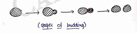
- It forms white, circular, smooth, creamy colonies on surface of media.
- They are aerobic, but some yeasts are facultative anaerobic.
- The optimum temp. for growth of yeast is .
- Yeast species are used in ethanol production and bakery industry. Examples : Saccharomyces cerevisiae, Cryptococcus neoformans etc...
iii) Yeast Like Fungi
In some yeast like Candida albicans, the bud remains attached to the mother cell and elongated, followed by repeated budding, forming chains of elongated cells known as pseudohyphae.
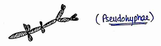
- It forms moist creamy coloured colonies on surface of solid media.
iv) Fungi Imperfectii
- Most of the species are used to production of enzymes and antibiotics.
- Examples : Aspergillus niger, Aspergillus fumigatus, Penicillium notatum etc-
- They are also called as Deuteromycetes or Hyphomycetes.
- They consists of group of fungi whose sexual phases are not identified.
- They grow as molds as well as yeasts.
- Most fungi of Medical importance belong to this class.
- Examples → Coccidioides immitis, Candida albicans.
iv) Dimorphic Fungi
Some fungi, mainly pathogenic species exhibit dimorphism i.e. two forms of growth.
- fungi can grow either as a mould or as a yeast.
- Dimorphism is temp. dependent. So, At it shows mould like growth and at it shows yeast like growth.
- Examples → Mucor rouxii, Histoplasma capsulatum etc...
Classification (Based on their sexual spore formation)
i) Zygomycetes
ii) Ascomycetes
iii) Basidiomycetes
iv) Deuteromycetes (Fungi imperfectii)
i) Zygomycetes
- They are fungi having non-septate hyphae (lower fungi).
- They form asexual spores (sporangiospore) contained within sac like structure called sporangia.
- Examples → Rhizopus, Mucor etc.
ii) Ascomycetes
- They form sexual spores within a sac and are called ascospores.
- sac is called ascus.
- They include both yeasts and moulds.
- Examples → Histoplasma, Candida etc...
iii) Basidiomycetes
They reproduce sexually and form septate hyphae.
- These basidiospores are borne at the tip of the basidium.
- Examples → Cryptococcus neoformans, mushrooms etc-
iii) Basidiomycetes
They reproduce sexually and form septate hyphae.
- These basidiospores are borne at the tip of the basidium.
- Examples → Cryptococcus neoformans, mushrooms etc-
Reproduction/Replication of fungi
- They reproduce by both sexually and asexually.
i) Sexual Reproduction of Fungi
In this reproduction, sexual spores is formed by fusion of two different cells (Male & female cell) fused called plasmogamy, then further fusion of two nuclei called Karyogamy.
- As a result zygote is formed, which further formed sexual spores.
- These are produced by meiosis.
- Like as Ascospores, Basidiospores, Zygospores.
ii) Asexual Reproduction of Fungi
In this reproduction, asexual spores are produced by mitosis.
i) Vegetative spores
ii) Aerial spores
i) Vegetative Spores
- Blastospores
- Arthrospores
- Chlamydospores
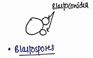
- Blastospores - These are formed by budding from parent cells, as in yeasts.
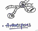
- Arthrospores :- These are formed by segmentation & condensation of hyphae.
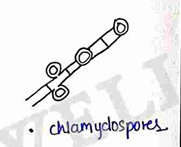
- Chlamydospores :- They are thick walled resting spores developed by rounding up and thickening of hyphal segments.
ii) Aerial Spores
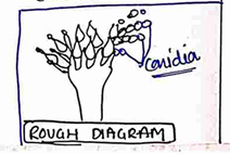
- Conidiospores : Spores borne externally on sides or tips of hyphal are called conidiospores or conidia.
Cultivation of Fungi
In cultivation of fungi, we have to identify fungi, then separate and grow fungi.
- Sabouraud agar is a type of agar growth medium containing peptones. It is used to cultivate dermatophytes and many types of fungi and also grow.
- It has utility for research and clinical care.
- It is developed by Raymond Sabouraud in 1892.
- The standard temp. for incubation of fungi is in humidified environment for 21 days.
- After 21 days colonies of fungi form on culture media.
- Media used for growth
- Brain-heart infusion (BHI) agar
- Czapek's agar etc-
VIRUSES
- Viruses are the smallest infectious agent, are very small, so we can watch it with the help of electron microscope (ultra microscope).
- They are about 10-100 times smaller than most of the bacteria.
- They are composed of genetic material DNA OR RNA (any one) and protein.
General structure of Viruses
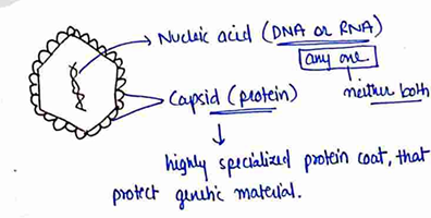
- Viruses are obligate parasites, i.e. totally depends on its host (they are incapable of independent growth in artificial media).
- Viruses are non-living thing until they enter inside the living body (host). So,
- Outside the living body (without host) Non-living
- Inside the living body (with host) Living thing.
- The complete infective form of a virus outside a host cell, with a core of RNA or DNA and a capsid (protein) is called Virions.
Virology → In which we study about viruses.
On the basis of their morphology, Viruses divided into three types
i) Helical virus (Rod shaped viruses)
ii) Polyhedral (Icosahedral) viruses.
iii) Complex virus
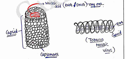
i) Helical Viruses
- They are rod-shaped like structure.
- They have capsid made from over thousand molecules of single type of protein arranged in a helix.
- The genetic material is generally single-stranded RNA (sometimes DNA) bound into the protein helix by interaction between the negatively charged nucleic acid and positive charge on protein.
- Eg. Rabies viruses, Tobacco Mosaic virus (TMV).
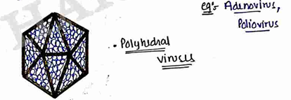
ii) Polyhedral (Icosahedral) Viruses
They are round shaped.
- Also called as spherical viruses.
- Icosahedral means, it have 20 sided/faces and may be more than 20 (polyhedral).
- It gives roughly spherical appearance.
- Eg. Adenovirus, Poliovirus.
iii) Complex Viruses
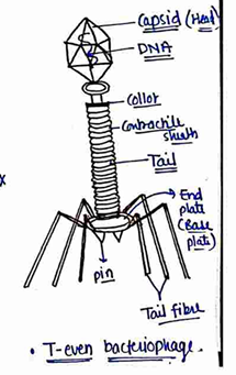
They are tad pole like structure i.e. (head and tail).
- It have very complicated structure, so it is called complex viruses.
- Eg. Bacteriophage virus.
Classification On the Basis of Host
i) Animal viruses
ii) Plant viruses
iii) Bacterial viruses (Bacteriophages)
i) Animal Viruses
They parasite on animals and Cause disease on them.
- They contain both DNA OR RNA (any one) as genetic material.
- They causes diseases → Polio, Hepatitis, AIDS or many more etc...
ii) Plant Viruses
They parasites on (infect) plants may be potato, sugarcane, tobacco etc and Cause disease on them.
- They have only RNA.
- Disease → Tobacco mosaic virus (TMV) etc...
iii) Bacteriophages
- The viruses infect on bacteria and Cause disease on bacteria.
- They contain only DNA as genetic material.
- They may be infect E.coli bacteria, and it is called T-even phages ($T_2, T_4, T_6$).
Replication/Reproduction of Viruses
For replication of virus host is necessary. host may be bacteria, plants or an animals.
When virus infect on host cell, a host cell is forced to rapidly produce thousands of identical copies of original virus.
Steps for Replication
i) Attachment
ii) Penetration
iii) Uncoating (Biosynthesis)
iv) Assembly (Maturation)
v) Release
i) Attachment
In this step, virus attached on the surface of host cell, they attached on specific receptor on plasma membrane of host cells.
ii) Penetration
In this step, virus enter inside the host cells, (only DNA/RNA of virus enter or complete virus enter). Entry of virus.
iii) Uncoating & Biosynthesis
In this step, viral Capsid is removed by viral enzymes or host enzyme.
- Due to capsid removed viral genomic nucleic acid release in host cells.
Biosynthesis (Genome replication) : The Replication mechanism depends on the viral genome.
- DNA viruses usually use host cell protein and enzymes to make additional DNA that is transcribed to messenger RNA, which is then used to direct protein synthesis.
- RNA viruses usually use the RNA core as a template for synthesis of viral genomic RNA.
iv) Assembly (Maturation)
After biosynthesis modified viral protein are packaged with newly replicated viral genome into new virions that are ready for release from the host cells.
v) Release
Two method for release:-
i) Lysis → In this viruses burst the cell membrane of host cell and release. (death of host cell). (Cytolytic).
ii) Budding → In this viruses, make envelope from plasma membrane of host cell and release, does not harm host cell. (Influenza).
Cultivation of Viruses
Viruses are obligate parasites, so they cannot grow on culture media.
- Cultivation of viruses are as follow :-
i) Laboratory Animals
It is one of the oldest method for cultivation of viruses.
- Animals use for this, such as rabbits, guinea pigs, mice (Rat), ferret etc-
- The growth of the virus in inoculated animals may be indicated by death, disease or visible lesions.
- Animal inoculation is used for the study of pathogenesis, immune response and epidemiology.
ii) Embryonated Eggs
The embryonated hen's egg offer several sites for the cultivation of viruses.
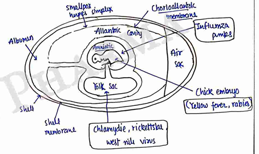
- Fertile chicken eggs incubated for 5-12 days can be inoculated through the shell aseptically. The opening may be sealed with paraffin wax and the egg incubated at for the time required for the growth of the virus.
- Viruses may kill the chick embryo and produce specific evidence of viral activity.
- This effect help in the identification of virus.
Classification and Mode of Action of Disinfectants
Disinfectants
Disinfectants are antimicrobial agents that are applied to non-living object to destroy microorganism, and the process is known as disinfection.
- It is not necessary that it kill all microorganism but reducing them to a level acceptable for a defined purpose. (a level which is harmful neither to health nor to the quality of perishable goods).
- Disinfectants are usually bacteriocidal but occasionally they may be bacteriostatic.
An ideal disinfectant should have the following properties:
i) Broad Spectrum
ii) Non Toxic
iii) Economical
iv) Odourless
v) Easy to Use
vi) Stable on StorageChemical agents are most commonly used as disinfectants. These chemical agents are classified as follow
i) Acids and Alkalies
ii) Halogens (imp)*
iii) Heavy Metals
iv) Phenol and Its Derivatives
v) Alcohols
vi) Aldehydes
vii) Dyes
viii) Detergents and Soaps
ix) Quaternary Ammonium Compounds
i) Acids and Alkalies
The germicidal efficiency of acids is proportional to the hydrogen ($H^+$) ion concentrations of their solutions.
- They produce toxic effects which decrease or kill bacteria (microorganism).
- Eg. Hydrochloric acid, Sulphuric acid, Nitric acid etc.
- Similarly, the germicidal action of alkalies is dependent on dissociation and release of hydroxyl ion ($OH^-$) in solution.
ii) Halogens
The germicidal action of halogens and its compounds is due to formation of hypochlorous acid (or other acids) when free halogens reacts with water.
Eg. Chlorine, Iodine, Bromine, flourine act as germicidal.
- The released in this reaction is a strong oxidising agent, it acts on the cellular constituents of microorganism.
- A combination of chlorine and protein of the cell membrane and enzyme is also responsible for the death of microorganism.
iii) Heavy Metals
The most widely used compounds of heavy metal are those of Mercury, silver and copper.
Heavy metals and their compounds act antimicrobials by combining with cellular proteins.
(active enzyme) (Inactive enzyme)
iv) Phenol and Its Derivatives
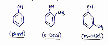
Phenol and its compounds mainly destroy the microorganism by the process of disruption of cells, precipitation of cell protein, inactivation of enzymes and leakage of amino acids from the cells.
- Some important phenols are
v) Alcohols
Alcohols are protein denaturants. They may damage lipid complexes in the cell membrane. They are also dehydration agents.
- Alcohol concentration above 60% are more effective against viruses.
- Alcohol are used as preservatives in some vaccines.
- Eg. Methanol ($CH_3OH$), Ethanol ($C_2H_5OH$), Isopropanol ($CH_3CH(OH)CH_3$), n-butanol etc....
vi) Aldehydes
Formaldehyde (HCHO) is the main aldehyde used for disinfection.
- It is used in the medical field for sterilizing lenses and other instrument (gaseous sterilization).
vii) Dyes
They inhibit bacterial growth. Basic dyes are more effective bactericides than acidic dyes.
- They impair the DNA complexes of microorganism and destroy the reproductive capacity of the cell.
- Used for the treatement of both burns, bladder irrigation.
- Eg : Acridine, triphenyl methane etc.-
viii) Detergents and Soaps
They are widely used surface active agents (surfactants).
- The most important antimicrobial agents are the cationic active agents. (quaternary ammonium compounds).
- Soaps and sodium lauryl sulfate are anionic compounds.
ix) Quaternary Ammonium Compounds
Most of the cationic detergents are quaternary ammonium compounds.
- They are highly active against Gram-positive bacteria and are also quite active against Gram-negative bacteria.
- Their mode of action is disruption of cell walls and membrane. They also inactive enzymes and denature proteins and thus kill a microorganism.
General structure
- Used on floors, used as skin antiseptics etc.-
Factor Affecting/Influencing Disinfectants
There are some following factors, which affect the action of disinfectants
i) Effect of Temperature
ii) Effect of pH
iii) Effect of Dilution
iv) Effect of Surface Activity
v) Effect of Interfering Agent
i) Effect of Temperature
If the temperature of the disinfectants is increased, the effectiveness of the disinfectants also increases.
ii) Effect of pH
The optimal bacterial growth take place in the range of 6-8, on either side of this the rate of growth declines.
- If we provide some pH, which bacteria need to grow, then bacteria grow and disinfectants became less effective.
- So, always provide different pH disinfectants to decline the growth of bacteria (microorganism).
iii) Effect of Dilution/Concentration
At higher concentration the drugs acts as bactericidal, while on dilution the drug act as bacteriostatic.
So, Concentration of disinfectant = effectiveness
- But also used in in limit.
iv) Effect of Surface Activity
The addition of low concentration of surface active compounds may potentiate the biological effect of an antibacterial agent.
Eg. Phenols are more active in the presence of soaps.
v) Effect of Interfering Agents
Interfering agents like organic acids, may directly or indirectly affect the action of disinfectants.
These interfering agents reduce the activity of disinfectants by adsorbing or inactivating them.
Evaluation of Disinfectants
There are various methods being employed to evaluate the potency of the disinfectants, following :-
1) Tube Dilution and Agar Plate Method
The chemical agent is incorporated into nutrient broth or agar medium and inoculated with the test microorganisms.
These tubes are incubated at for 2-3 days and then the results in the form of turbidity or colonies are observed.
 a) Tube dilution method
 b) Agar plate dilution method
- The results are recorded and the activity of the given disinfectant is compared as shown in fig .
2) Cup-plate Method
In these method, the agar is melted, cooled at about and inoculated with the test microorganism and poured into a sterile petri plate.
- When the inoculated agar has solidified, holes about 9mm in diameter are cut in the medium with a sterile cork borer.
- The antimicrobial agent is directly placed in the holes and the zone of inhibition is observed after incubation at 30 to for 2-3 days.
- The diameter of the zone of inhibition gives an indication of the relative activities of different antimicrobial substances against the test microorganisms.
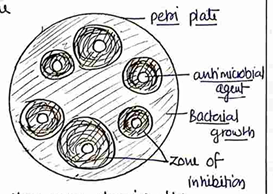
- Clear zone showing the no growth of bacteria.
3) Phenol Coefficient Method
In this method, a test chemical is assayed for its microbicidal property with reference to phenol under identical conditions.
- Organisms used in these methods are :- Salmonella typhi, Staphylococcus aureus, Pseudomonas aeruginosa are used.
Phenol coefficient test includes :
i) Rideal-walker test (RW test)
ii) Chick-Martin test (CM test)
- The phenol coefficient of test disinfectants may be calculated by Rideal-walker test that use Rideal-walker broth and Salmonella typhi as the sensitive microorganism.
Procedure
- Different dilutions of the test disinfectant and phenol are prepared and 5ml of each dilutions is inoculated with 0.5 ml of the 24 hours broth culture of the organisms.
- All tubes (disinfectants + organism and phenol + organism) are placed in a water bath.
- Subcultures of each reaction mixture are taken and transferred to 5ml sterile broth after 2.5, 5, 7.5 and 10 min.
- The both tubes are incubated at for 48 to 72 hours and are examined for the presence or absence of growth.
- Result show in table . (+) = it show growth (-) = it show no growth.
| Disinfectant | Dilution | Time interval for sub-culture (min.) 2.5 | 5 | 7.5 | 10 |
|---|---|---|---|---|---|
| Test disinfectant | 1:1000 | + | - | - | - |
| 1:2000 | + | + | - | - | |
| 1:3000 | + | + | + | - | |
| 1:4000 | + | + | + | + | |
| Phenol | 1:80 | + | - | - | - |
| 1:100 | + | + | - | - | |
| 1:120 | + | + | + | - | |
| 1:140 | + | + | + | + |
Now, Calculated Rideal-walker coefficient of the disinfectants
If the phenol coefficient (RW coefficient) of given test disinfectant is :
1 = same effectiveness as phenol.
Less than 1 = less effective than phenol. More than 1 = More effective than phenol.
- Here is 20, means test disinfectant is 20 times more active than phenol.
Chick Martin Test
This type of test is done in controlled amount of organic matter in the form of standardization suspension of yeast cells.
In this test all the conditions like Rideal Walker test but the temperature is and exposure time is 30 minutes.
For Example :
| Tube | Test disinfectant % | Tube | Phenol % | ||
|---|---|---|---|---|---|
| 1 | 2.0 | - | 1 | 1.80 | - |
| 2 | 0.47 | - | 2 | 1.62 | + |
| 3 | 0.41 | + | 3 | 1.45 | + |
| 4 | 0.37 | + | 4 | 0.33 | + |
- Mean for the disinfectant Under test =
- Mean for Phenol =
- The chick Martin coefficient is the mean of the highest concentration of phenol showing growth and lowest concentration preventing growth, divided by the same mean for the disinfectant Under test. $$\text{Phenol coefficient} = \frac{1.625}{0.39} = 4.16$$
- Greater than one, so more effective (test disinfectants) than phenol disinfectants.
4) Kelsey-Sykes Test
It is a triple challenge test
(test disinfectant checked three times with successive addition of microbial suspension with the same concentration of disinfectants).
- This test is performed in both condition clean (no organic matter) as well as dirty (with organic matter).
- In this method several test bacteria such as Staphylococcus aureus, Proteus vulgaries, Escherichia coli and Pseudomonas aeruginosa are used.
- Dilution of disinfectant are made in hard water. The duration of test is 30 min.
Procedure
Inoculate 3ml of the disinfectants dilution with 1ml of bacterial suspension in broth (clean) OR yeast (dirty) and shake gently.
After 8 min, transfer 1 drop to each of 5 tubes OR a surface of nutrient agar plate, to check the disinfectants kill microbial cell or not.
Now, add 1ml of microbial cell into same mixture (disinfectant + bacterial suspension). After 10 min.
After 18 minutes from process starts, Again transfer microbial cell to nutrient media for check.
Again add 1ml of microbial cell into same mixture. After 10 min.
After 28 min from processing start, again transfer microbial cell to nutrient agar plate to check the disinfectants.
- After this, sample taken at 8, 18, 28 minutes are then incubated at and the number of tubes showing growth OR the number of colonies/drop from surface culture is recorded.
Interpretation of Result
The test disinfectants is reported as PASS if it kills the bacteria in at least three tubes/petri plates of the 18 minutes samples.
Sterility testing of Products (Solids, Liquids, Opthalmic and other sterile Products) according to IP, BP and USP.
- Action of disinfectants (Antimicrobial agents)
Bacteriostatic
These are those antimicrobial agent which inhibit the growth of microorganism. or
We say that, they stop the multiplication of microorganism (bacteria).
Bactericidal
These are those antimicrobial agent which are capable of killing microorganism (bacteria). or we say that, they stop the growth of bacteria by killing them.
- After the sterilization process of any product, material we have to check whether the product (material) that we have sterilized is fully free of bacteria (microorganism) or not.
- For this we use some test of sterility for that products, which is known as sterility testing of products.
- The tests for sterility are intended for detecting the presence of viable form of microorganism in pharmaceutical preparations.
- The test for sterility is applied according to IP, BP and USP.
- A number of products sterilized such as Ophthalmic preparations, injections, infusions, implants, syringes, bandages, dressing, needles, surgical instruments etc_
- Test must be carried out under aseptic condition designed to avoid accidental contamination of the drug during testing.
- For all these condition, Laminar air flow is recommended.
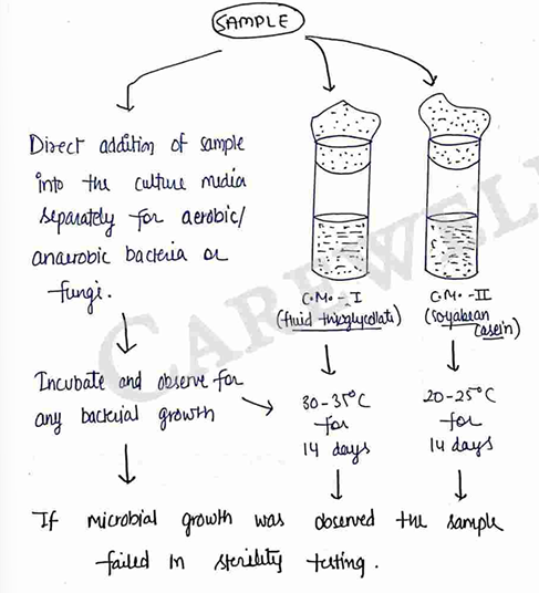
- Culture media designed for sterility testing.
- Fluid Thioglycollate → For anaerobic bacteria, also for some aerobic bacteria.
ii) Soyabean Casein Digest → For fungi and aerobic bacteria.
- Before performing test, also checked the suitability of media by
- Sterility testing of the media (C.M. pure)
- Growth promotion test (check the ability of culture media to grow bacteria)
Methods for Sterility Test
Sterility test can be carried out by using the following methods :-
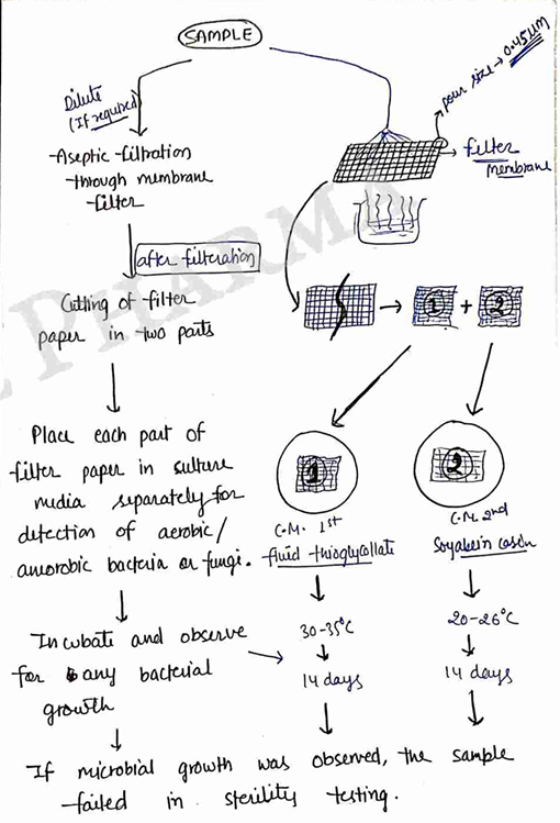
- Method A: Membrane filteration.
- Method B: Direct Inoculation.
1) Method A :- Membrane filteration
Products :- (an oil, ointment, soluble powder, liquid products etc_)
2) Method B - Direct Inoculation
Products (aqueous solution, suspension, oily soluble products, surgical dressing, sterile devices etc...)