Pathophysiology - Unit 1
Syllabus
Basic principles of Cell injury and Adaptation:{" "} Introduction, definitions, Homeostasis, Components and Types of Feedback systems, Causes of cellular injury,Pathogenesis (Cell membrane damage, Mitochondrial damage, Ribosome damage, Nuclear damage),Morphology of cell injury - Adaptive changes (Atrophy, Hypertrophy, hyperplasia, Metaplasia, Dysplasia),Cell swelling, Intra cellular accumulation, Calcification, Enzyme leakage and Cell Death Acidosis &Alkalosis,Electrolyte imbalance
Basic mechanism involved in the process of inflammation and repair: Introduction, Clinical signs of inflammation, Different types of Inflammation,Mechanism of Inflammation - Alteration in vascular permeability and blood flow, migration of WBC’s,Mediators of inflammation,Basic principles of wound healing in the skin,Pathophysiology of Atherosclerosis
Scroll to Download
PATHOPHYSIOLOGY (UNIT-1)
BASIC PRINCIPLES OF CELL INJURY AND ADAPTATION
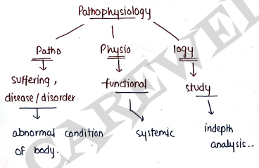
PATHOPHYSIOLOGY:
- It is the branch of science which deal with the study of disease/abnormality occured inside the body.
- It also involves the study of those changes which occurs in our body due to any disease.
- It involves the study of:-
- causes of disease
- Sign and symptoms
- change occured in body (Biological changes)
- functional changes
- complications, diagnosis and treatement.
Some Basic Terminology
- Disease It is the abnormal condition of body.
- Etiology It is the study about those factors which causes disease.
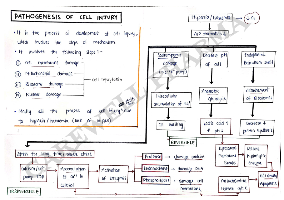
- Pathogenesis It involves the mechanism and steps involves in occurance of disease.
- Morphology It is the study of structure of cells.
- Sign & symptoms These are those changes, which occures due to disease.
SYLLABUS
Introduction, definitions of Homeostasis components and types of feedback system.
Causes of cell injury, pathogenesis (cell membrane damage, mitochondrial damage, Ribosome damage, Nuclear damage), Morphology of cell injury - Adaptive changes (Atrophy, Hypertrophy, Hyperplasia, Metaplasia, Dysplasia), cell swelling, Intra cellular accumulation, calcification, Enzyme leakage and cell death acidosis & alkalosis, Electrolyte imbalance.
Pathophysiology
It is the branch of science which deal with the study of disease and abnormal changes occured in body functions.
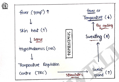
HOMEOSTASIS
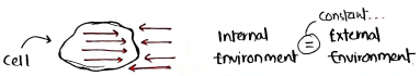
- It is a condition when our cell's internal environment is constant with respect to external environment.
- It is derived from two greek words:-
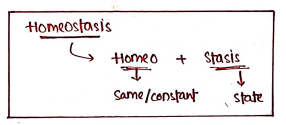
- It means staying the same/constant by maintaining pH, temperature, acid-base balance etc.
Components of feedback system
- All the body organs coordinate with each other to maintain homeostasis.
- This coordination/maintainence of homeostasis is controlled by feedback system with the help of Neuroendocrine (Nervous + Endocrine) system.
It has three components:-
- Receptors: It is a type of sensor which receive/detects changes or other stimuli.
- Control Centre: It receive the stimuli from receptors and analyse it.
- Effectors: It response to maintain homeostasis.
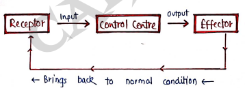
Feedback System: If there are any change take place in internal environment then feedback system is take back into its constant state or in homeostasis.
It is of two types:-
- Positive feedback system (+) ve
- Negative feedback system (-) ve
Positive feedback system (+) ve
- used to increase...
- When anything is decrease in our internal environment, then it is try to back into its normal situation by increasing it.
- eg. During childbirth, it stimulate the release of oxytocin which increases the contraction of the uterus to help in childbirth.
Negative feedback system (-) ve
- Used to decreases...
- When anything is increases in our internal environment (body), then this system is try to back into normal condition by decreasing it.
CELL INJURY
- Cells are the smallest structural and functional unit of body and they maintain their normal homeostasis.
- When a cell encounters a variety of stresses, their will be a changes in its internal as well as external environment leads to cell injury.
- Cell injury is defined as, it is change/alteration in cell structure or functions due to some stress that exceed the ability of cells to handle it.
- When injury is severe, it may lead to cell death.
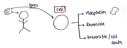
- Cells may respond in three way to any stress.
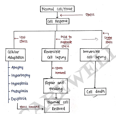
- Cellular Adaptations : It is a condition when cells adjust (adapt) themselves with the changing environment when exposed to stress, to survive and maintain the normal state i.e. homeostasis.
- It is occured due to functional demand and revert back to its normal state when stress removed.
eg. Atrophy, Hypertrophy etc...
Reversible cell Injury : It is a condition when mild to moderate stress is applied and cell back to its normal state if stress is removed through repair & healing.
Irreversible cell Injury : It is occured when cells are exposed to severe stress or continuous stress. In this condition, cells never revert back and leads to cell death.
eg. Apoptosis, Necrosis.
CAUSES OF CELL INJURY (Etiology)
- The causes for both reversible and irreversible cell injury are similar:-
- Hypoxia and Ischaemia
- Physical Agents
- Immunological Agents
- Chemicals and Drugs
- Microbial Agents
- Psychological Factors
- Nutritional Balance
- Genetics Dearrangements
Hypoxia and Ischaemia :
- Hypoxia is the one of the most important cause of cell injury.
- Hypoxia is the condition of loss of oxygen supply in body/cells which starts the process of cell injury, because oxygen is most important for various metabolic functions and generation of energy/ATP.
- Ischaemia is the condition of low supply of blood which leads to hypoxia, because oxygen is transported through blood.
Physical Agents :
- Various physical agents are responsible for cell injury.
Eg. Trauma (Road accident), Heat, Cold, Electricity, Radiations (UV light, sun burn) and rapid change in atmosphere etc.
- Various physical agents are responsible for cell injury.
Immunological Agents :
- Immune system serves as one of the defence against bacteria / injurious agents, but sometimes it also cause cell injury.
Eg. Hypersensitivity reactions, Autoimmune disease, Anaphylactic reactions etc...
- Immune system serves as one of the defence against bacteria / injurious agents, but sometimes it also cause cell injury.
Chemicals and Drugs:
- Various chemicals and drugs may cause cell injury.
Eg. chemically poisonous substances Cyanide, arsenic, mercury etc.
High oxygen concentration.
Glucose and salt in hypertonic solutions.
Drug Abuse/overdose etc.
- Various chemicals and drugs may cause cell injury.
Microbial Agents:
- Various microbes such as bacteria, viruses, fungi, protozoa etc may cause infection/cell injury.
Eg. Dengue is caused by flavi virus. Corona virus etc...
- Various microbes such as bacteria, viruses, fungi, protozoa etc may cause infection/cell injury.
Psychological factors:
- Various mental disorder/addiction such as alcoholism, drug addiction and smoking results in disease such as liver damage, lung cancer, peptic ulcer and Cardiovascular disease.
Nutritional balance:
- Sometimes excess or deficiency of nutrients may cause cell injury.
Eg. Deficiency of proteins (kwashiorkor disease),
Deficiency of minerals (anaemia),
Excess of lipids (obesity and atherosclerosis).
- Sometimes excess or deficiency of nutrients may cause cell injury.
Genetic factors:
- sometimes cell injury occurs due to defects in genes or chromosomes. Sometimes some disease/cell injury occurs in child from parents through heredity.
eg. Teratogenes (responcible for birth defects), sickle cell anaemia, diabetes mellitus, Hypertension etc..
- sometimes cell injury occurs due to defects in genes or chromosomes. Sometimes some disease/cell injury occurs in child from parents through heredity.
MORPHOLOGY OF CELL INJURY
- Morphology is defined as it is the study of structure and forms of cells.
- It involves the cellular adaptation when cell experienced minimal stress and due to this adaptive changes take place in cells.
CELLULAR ADAPTATION
- These are adaptive changes which occurs when cells adjust (adapt) themselves with the changing environment when exposed to stress, to survive and maintain the normal homeostate.
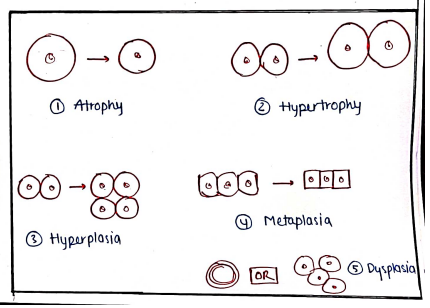
It is of mainly five types:-
- Atrophy
- Hyperhophy
- Hyperplosia
- Metoplasia
- Dysplasia
Atrophy:
- It is defined as it is the decrease/reduction in the cell size which may also reduce its functions.
- It may caused due to poor nutrition, reduced blood supply, loss of endocrine stimulation (i.e. hormones) and ageing.
- It can be physiological (eg. loss of hormone, ageing) and pathological (eg. starvation, ischemia etc).
Hypertrophy:
- It is defined as it is the increase in the size of the cells.
- It may caused due to increase in the no. of organelles and amount of structural proteins.
- It occured due to increase in functional demand or stimulation by specific hormones.
- eg. enlarged size of uterus during pregnancy (physiology), hypertrophy of cardiac, smooth muscles (Pathological).
Hyperplasia:
- It is defined as it is increase in the number of cells.
- It is occured due to increases cell replication and it is accompanied with hypertrophy, in response to same stimuli.
Eg. overproduction of hormones and growth factors.
Metaplasia:
- It is defined as it is the reversible change in the types of cells.
- In this one type of cell is converted into another and its function can also be changed.
- It can be caused due to prolonged irritation, hormone disturbance, nutritional deficiency & infections.
eg. Columnar epithelial converts into squamous epithelial.
Dysplasia:
- atypical hyperplasia.
- It is defined as it is the abnormal or disordered development of cells or irregular growth.
- It occurs along with metaplasia & hyperplasia.
- It occurs due to chronic irritation and prolonged inflammation.
eg. increase in no. of layers of epithelial cells. arrangement of cells in disorganised manner. etc..
FOR REVERSIBLE CELL INJURY
- It is a condition/injury which occurs when mild to moderate stress is applied and in this cell back to its normal state if stress is removed.
It is mainly of four types:-
- Hydropic Changes
- Hyaline Changes
- Mucoid Changes
- Fatty Changes
Hydropic changes:
- It is also know as cell swelling.
- Cellular swelling occurs due to accumulation of extracellular ions and water inside the cell.
- During injury, Hypoxia/ischemia occurs which stops the Na^+$/$K^+ pump.
- It leads to accumulation of in cell.
- Now, water and other ions enters into cell to maintain iso-osmotic pressure which leads to cell swelling. (Intracellular accumulation) along with .
Hyaline changes:
- It is defined as it is glassy, homogeneous, appearance of cells.
- It is occured due to accumulation of plasma proteins or other proteins.
- It mainly occured on the sites of epithelial and connective tissue.
eg. Russell's Bodies, Hyaline droplets, Benign tumor etc. (immunoglobulins).
Mucoid changes:
It is defined as it is the accumulation of mucin (part of mucus) in cells.
It is of two types:-
- Epithelial mucin - eg. cystic fibrosis of pancrease
- Connective tissue mucin - eg. mucoid in some tumors (mesenchymal tumor).
Fatty changes:
- Also known as Steatosis.
- It is defined as it is the abnormal accumulation of triglycerides/fat in cells.
- It is caused by toxins, diabetes, obesity, alcohol abuse, protein malnutrition etc..
- It mainly occurs in liver (eg. fatty liver) and also in heart, skeletal muscles etc..
INFLAMMATION
CHAPTER-2 UNIT-1
SYLLABUS
Basic mechanism involved in the process of inflammation and repair: Introduction, clinical sing of inflammation, Different types of inflammation.
Mechanism of inflammation - Alteration in vascular permeability and blood flow, Migration of WBC's, Mediators of inflammation.
Basic principles of wound healing in the skin, pathophysiology of Atherosclerosis.
- Inflammation is defined as it is the local response of our body to an injury occured from any type of agents.
- These are protective responses of our body's immune system in which leukocytes (WBCs) transported to injured site and fight against agents (bacteria etc) and eliminated or limited.
- It is our body's defense mechanism.
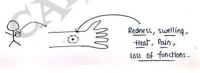
Clinical sign of inflammation
These are the sign/response of inflammation:-
Rubor (Redness): Redness at the site of inflammation due to vasodilation.
Tumour (Swelling): swelling at the site of inflammation due to exudation.
Calor (Heat): elevated temperature at the site of inflammation due to increased blood flow.
Dolor (Pain): pain at the site of inflammation due to release of chemical mediators.
Loss of functions: The cells at the site of inflammation does not work/function properly.
Causes of Inflammation
These are those agents which are responsible for inflammation:-
- Physical Agents: Heat, cold, radiations, trauma etc..
- Chemical Agents: Chemicals, drugs, toxins, poison etc..
- Biological Agents: Bacteria, Virus, other microbes.
- Immunological Agents: Hypersensitivity reactions, antigen-antibody reactions etc.
TYPES OF INFLAMMATION
On the basis of time of duration, it can be classified into two types:
- Acute Inflammation
- Chronic Inflammation
Acute inflammation:
- It occurs for short time period and show the early body reactions.
- It is an immediate or quick response to sudden body damage (infection, trauma etc)
- It is beneficial for our body and provide protective response against injury.
eg. fever, pain, Redness, swelling etc..
Chronic inflammation:
- It occurs for a longer time period when the causative agents of acute inflammation remains for a longer duration.
- It is also referred to as slow, long-term inflammation lasting for prolonged periods of time i.e. several months to year.
eg. Asthma, Alzheimer's disease, peptic ulcer etc..
MECHANISM OF INFLAMMATION
It is the pathogenesis of inflammation which includes the steps involves in the mechanism of inflammation.
It includes:
I. Vascular events
II. Cellular events
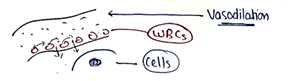
I. Vascular events
These are the early responses occured after any type of injury in blood vessels.
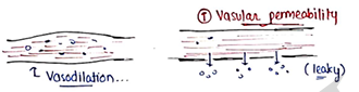
- It involves changes/alteration in vascular permeability and changes in blood flow.
- It involves the vasodilation of blood vessels which increases the blood flow at the site of inflammation.
- It increases the vascular permeability of blood vessels which helps in the migration of WBC's at the site of inflammation.
II. Cellular events
These are those responses which occured inside cells.
It involves several steps:
1. Migration of WBCs
- Initially when cells experienced any injury, vasodilation occurs at nearest blood vessels which increases the blood flow and amount of blood.
- Now, WBCs (most cells, neutrophills etc.) are agrregated in blood vessels at the site of inflammation.
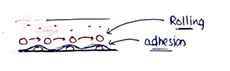
2. Rolling and adhesion
- Now WBCs roll order the endothelial cells slowly and this termed as rolling.
- Then, cell adhesion molecules like selections immunoglobulins, integrins binds WBCs (leukocytes) to the endothelial cells and this termed as adhesion.
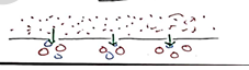
3. Emigration
- When WBCs stick to the endothelium, they search appropriate site.
- Then they leak into extracelluar space by damaging basement membrane by secretion of collagenoses enzyme or through pseudopods.
- Initially neutrophils then monocyte macrophages, and also some RBCs escape through gaps b/w endothelial cells (diapedesis)
4. Chemotaxis
- It involves the transmigration of ieukocytes (WBCs) at the site of inflammation (in cells) with the help of chemotoctis factors (eg. LT-B4, PF4, Cytokines etc..)
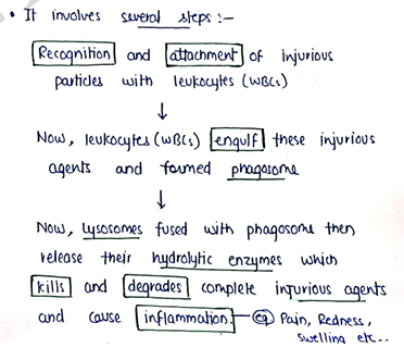
5. Phagocytosis
- It involves the process of killing or englufment of agents (that cause inflammation) which limits or eliminates the injurious agents.
Alteration in Vascular Permeability & Blood flow
- When cell experienced any type of injury, there will be increased in vascular permeability.
- This also increases the blood flow due to vasodilation.
- It involves following mechanism:-
- Contraction of Endothelial cells:
- It involves the contraction of endothelial cells, which forms gap b/w cells and increases vascular leakiness. Only in venules. Mediated by histamine, bradykinin etc.
- Retraction of Endothelial cells:
- It involves the contraction of endothelial cells, which forms gap b/w cells and increases vascular leakiness.

- Direct injury to endothelial cells:
- It involves direct injury to cells due to any injury/trauma which increases leakiness.
- It occurs in capillaries, venules and arteriols.
Mediated by leukocytes: At the site of inflammation, leukocytes (WBCs) activates and also increase vascular leakiness by releasing some mediators/enzymes.
Neovascularisation: Acc. to this, newly formed capillaries under Vascular Endothelial Growth factors (VEGF) are very leaky.
Haemodynamic Changes:
It involves the alteration in blood flow i.e. increases in the blood flow.
It involves several steps:
- Initially there is vasoconstriction (about in seconds)
- then persistent progressive vasodilation
- then WBCs follow emigration.
- Vasodilation increases the blood flow, appears red and little warm at site of inflammation.
It can also defined by Triple Response:
- Red line: It involve redness/redline on the skin occurs due to vasodilation.
- Flare: It involve appearance of flush (a bright reddish appearance) around redline.
- Wheal: It involve the swelling/oedema of the skin occurs due to transudation of WBCs/fluid into extracellular space.
BASIC PRINCIPLES OF WOUND HEALING IN THE SKIN
Wound
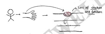
- It is an injury which is caused by any type of stress/force which break or cut the continuity of skin/tissue.
- It can also caused by any type of infection bacteria/virus. Leads to loss of structure and functions.
Healing
- It is the body's response to an injury in order to restore normal structure and functions. It involves two processes:
Regeneration: It involves complete restoration of original tissue by proliferation of parenchymal cells.
Repair: It also involves restoration of tissue but it result in the formation of scar.
Wound Healing: It is defined as it is the repair and regeneration of damaged tissue occured due to any type of injury.
Phases of wound healing
It involves four phases:-
- Hemostasis
- Inflammation
- Proliferation
- Maturation/Remodeling
- Hemostasis :
- It is the first step of healing.
- It activates the blood clotting system immiadate after an injury to stop bleeding and prevent blood loss.
- It involves vasoconstriction at the site of injury which follow up by various clotting factor and leads to formation of clots.
- Inflammation :
- It is the second step of healing.
- It involves migration of WBCs/Leukocytes at the site of injury which limit or eliminates the injurious agents.
- It increases vascular permeability, local release of cytokines and growth factors, macrophages (destroy bacteria & clear debris).
- At last, it shows the inflammatory responses such as pain, swelling, redness, heat etc.
- Proliferation:
- It is the third step of healing.
- It involves the regeneration and repair of wounds by proliferation of cells from surrounding site.
- Fibroblasts migrates into wound site and proliferates and Endothelial cells forms (new) capillaries by angiogenesis.
- It involves filling the wounds, contraction of wound margins then covering the wounds.
- Maturation/Remodelling:
- It is the fourth and final step of healing.
- The main feature of the maturation phase is the deposition of collagen in the wound.
- It involves remodelling of tissue and overall strength of tissue increased.
- It lasts from 21 days - 2 years depends on types of wound [i.e. first intention or second intention].
Types of Wound Healing
It is of two types:-
- Healing by first intention (Primary Union)
- Healing by second intention (secondary union)
Healing by first intention (Primary Union):
In this, wound should be:
- clean and free of infection
- Surgically cut
- Should not have lost much cells and tissue
- Wound edges surgically stiched.
It involves several steps:
- Initial haemorrhage
- Acute inflammatory responses
- Epithelial changes
- Organisation
- Suture tracks.
Healing by second intention (secondary union):
In this, wound should be:
- Open and infected with a large tissue defect
- With much loss of cells and tissues
- Wound edges left unstiched
It involves several steps:
- Initial haemorrhage
- Inflammatory phase
- Epithelial change
- Granulation tissue/proliferation
- Wound contraction.
factor affecting Healing
Local factors:
- Infection
- Poor blood supply
- Foreign bodies
- Type, Size and location.
Systemic factors:
- Age
- Nutrition
- Systemic infection
- Disease