Human Anatomy And Physiology 2 - Unit 3
Syllabus
Respiratory system
Anatomy of respiratory system with special reference to anatomy of lungs, mechanism of respiration, regulation of respiration
Lung Volumes and capacities transport of respiratory gases, artificial respiration, and resuscitation methods.
Urinary system Anatomy of urinary tract with special reference to anatomy of kidney and nephrons, functions of kidney and urinary tract, physiology of urine formation, micturition reflex and role of kidneys in acid base balance, role of RAS in kidney and disorders of kidney.
Scroll to Download
RESPIRATORY SYSTEM
CHAPTER-1
UNIT-3
SYLLABUS + IMPORTANT QUESTIONS
Anatomy of respiratory system with special reference to anatomy of lungs, Mechanism of respiration, regulation of respiration, Lung Volumes and Capacities, transport of respiratory gases, artificial respiration and resuscitation methods.
INTRODUCTION
- Respiratory system is a group of organs and tissues which help us in breathing or Respiration.
- These group of organs/tissue are known as Respiratory tract which includes the passage of air from the Nose to the alveoli in the lungs.
- Breathing It is generally a physical process of taking air into the body/lungs and expelling it. (mechanical)
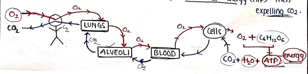
- Respiration It is the complete process of exchanging gases ($O_2$/$CO_2$) which includes Breathing (getting air into lungs), transport of gases to the body's cells and production of energy (ATP) then expelling .

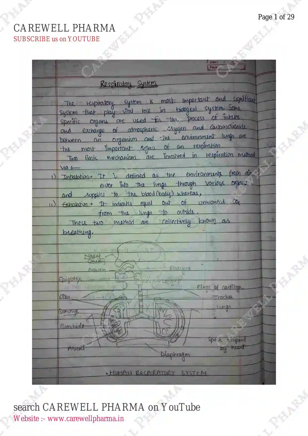
ANATOMY OF RESPIRATORY SYSTEM
The respiratory system consist of following Organs:
Upper Respiratory tract:
- Nose/Nasal Cavity
- Mouth / Oral cavity
- Pharynx
- Larynx
Lower Respiratory tract:
- Trachea
- Bronchi
- Bronchioles
- Lungs & Alveoli
- Diaphragm
- Coastal muscle

1) Nasal cavity (Nose)
The nose is the external organ of respiratory system responsible for inhaling & exhaling air.

Anatomy It includes:-
- External Nose: Visible part of Nose
- Nasal Cavity: internal passage of nose
- Nostrils: the opening of the Nose.
- Septum: thin wall separating two nasal passage.
- Turbinate bones: these are curved bones that warm and humidify air.
Physiology/functions:
- Air enters through nostrils.
- Air is warmed, humidified (moisture) and filtered with the help of hairs.
- It contains olfactory receptors for smell.
2) Mouth
It is not considered as a part of respiratory, but it is used as an alternate entry pathway for air (when nose is congested).
Anatomy
- It includes Oral Cavity, Lips, Tongue, palate (roof of mouth).
Physiology
- It is used for breathing/entry of air.
- It helps in chewing & contains taste buds for taste.
3) Pharynx
It is a muscular tube that connects the nose and mouth to the larynx (voice box).
- It is a common pathway for respiratory and digestive system.

Anatomy
- It is about long.
- It includes:
- Nasopharynx (upper part)
- Oropharynx (middle part)
- Laryngopharynx (lower part)
Functions
- It work as passage for air and food.
- It contains lymphoid tissue (tonsils, adenoids) help fight infections.
4) Larynx (Voice Box)
It is a cartilaginous organ responsible for producing sound. 
Anatomy
- present in the anterior neck at the 3rd and 6th cervical vertebrae level.
- A prominent elevation called Adam's apple is present in front of larynx.
- It includes thyroid cartilage (main), Cricoid (ring-shaped), Arytenoid (small cartilage) and vocal cords.
Physiology
- It produced sound through vocal cord vibration, also pitch and volume are regulated.
- It passes air from pharynx to trachea.
5) Trachea
It is also known as wind pipe. It connects the larynx to the bronchi.

Anatomy
- It is long.
- It is composed of 16-20 C-shaped rings of hyaline cartilage lying one above another.
- It is made up from cricoid cartilage (ring-shaped cartilage) & tracheal rings.
Physiology
- It passes the air into lungs.
- It keeps airway open.
- It also warms, humidifies and filters the inhaled air.
- It also generate cough reflex.
6) Bronchi and Bronchioles
- Bronchi are tubes that branch off from the trachea.
- Bronchioles are small tubes that branch off from the bronchi.
- These are simply a part of trachea.
- Bronchi: passage for air.
- Bronchioles: It leads to alveoli (passes air into alveoli for gas exchange).
7) Alveoli
These are tiny air sacs (hollow) where exchange of gases occurs with blood.

Anatomy
- A human lung has around 300 Million alveoli, covered with capillaries (70% area).
- Type 1 Alveolar cells (squamous): thin cells for gas exchange (around 95%).
- Type 2 Alveolar cells (cuboidal): thicker cell produce surfactants.
Physiology
- It exchange oxygen and carbon dioxide.
- Oxygen is absorbed into the blood from alveoli and is removed from the blood into alveoli.
- Surfactant helps to expand and deflate.
LUNGS
A pair of cone-shaped, spongy organs located in the thoracic cavity, responsible for exchanging oxygen & carbon dioxide through respiration.
- They are the primary organs of the respiratory system.

Anatomy:
- present within thoracic cavity (i.e. left lung & right lungs on each side of heart).
- Right lung is slightly larger than left.
- Weight (in adults): Right (approx. 600-700 gm), left (approx. 500-600 gm). Total .
- Right lungs have three lobes & left have two lobes.
Parts:
- Apex (upper facing)
- Base (concave/semilunar lower facing)
- Costal surface (costal cartilages, ribs and intercostal muscles).
- Medial surface (concave medial surface, contain triangular-shaped hilum - entry point for all).
- Mediastinum area present b/w lungs, which is occupied by the heart.
Layers:
- Each lung is enclosed within a pleural membrane.
- Pleural membrane: Parietal pleura (outer layer) & Visceral pleura (inner/deep layer).
- Pleural cavity (space b/w pleural membrane) contains pleural fluids helps to prevent lungs, shock absorbents and it allow membrane to glide over one another during breathing.
Lobes:
- Right lungs: 3 lobes (Superior, Middle, Inferior lobe)
- Left lungs: 2 lobes (Superior, Inferior)
- Each lobe is further divided into lobules, which contain alveoli.
Inner lungs: Inside, lungs contains:-
- Bronchi and bronchioles
- Alveoli which helps in respiration.
Diaphragm:
It is a dome-shaped muscles that separates the chest cavity from the abdominal cavity.
- It helps in contractions and relaxation of lungs to help breath in and out.
- helps to increase lung volume during inhalation.
functions/physiology
- Respiration: responsible for exchanging oxygen and .
- Filteration: lungs also filter out dust, bacteria and other particles from the inhaled air.
- pH level: lungs helps to regulate pH levels by removing excess ions.
MECHANISM OF RESPIRATION
Respiration : It is the process of exchanging oxygen and carbon dioxide between the body and environment. It involves the coordination of multiple organs and system, including the lungs, diaphragm, rib cage and nervous system.
- Environment (O_2$/$CO_2$) $\rightleftharpoons Body ($CO_2$/$O_2$)
- The average breathing rate of an adult is 15-18 times per minute (upto 25 times in childrens).
It involves several steps:-
- Inhalation/Inspiration
- Exhalation/Expiration
- Gaseous exchange
1) Inhalation/Inspiration
- process of intake of oxygen/air into lungs from environment (active process, required energy).
- It is the entry of air in lungs (entry of Oxygen $O_2$).
- It involves:-
- Diaphragm contracts and flattens, increasing lung capacity.
- Rib cage expands, intercostal muscle contract, expanding rib cage.
- Air enters nostrils or mouth, passes through pharynx & larynx.
- Epiglottis closes, directly air into trachea.
- Oxygen reaches to alveoli.
2) Exhalation / Expiration
- It is the process of removal of Carbon dioxide from lungs to the environment.
- It involves:-
- Diaphragm relaxes and domes, decreasing thoracic cavity.
- Intercostal muscles relax, decreasing rib cage size.
- Air leaves alveoli.
- Epiglottis opens and air exits through nostrils or mouth.

3) Gaseous Exchange
- It is the process of exchanging of and between Alveoli of lungs and blood.
- It occurs between inhalation and exhalation.

- It includes the following mechanism:-
- Oxygen diffuse from alveoli into blood through alveolar-capillary membrane by the mechanism of diffusion (High pressure to low).
- diffuse into alveoli by same way.
- transported to blood and then to cells through same diffusion mechanism and exhaled out from body. (Cellular Respiration)
REGULATION OF RESPIRATION
- The average rate of respiration is about 15-18 per minutes, more in childrens.
- Regulated by:- Nervous System & chemical control of some factors.
Nervous system -
- The voluntary control centre located in the cerebral cortex, sends impulse to the respiratory motor neurons via corticospinal tract.
- Automatic control:
- Medulla oblongata: primary respiratory centre, controls breathing rate & depth.
- Pons: secondary respiratory centre, regulate breathing rhythm.
- Chemoreceptors: detects change in oxygen and level & send signals to respiratory centres.
- Stretch receptors: detect change in lung volume, send signal to centers.
Chemical Control -
- These are depends on the amount of:
- Carbon dioxide ($CO_2$)
- Oxygen ($O_2$)
- also depend on the pH.
Other factors -
- Exercise ($\uparrow$ breathing rate, due to in the demands)
- Emotions
- Altitudes
- Temperature
LUNG VOLUMES
These refers to the amount of air in the lungs during different stages of breathing.
- Tidal Volume (TV) : The amount of air inhaled during normal breathing. Approximately .
- Inspiratory reserve Volume (IRV): It is the additional air that can be inhaled after normal inhalation. Approx. .
- Expiratory reserve volume (ERV): It is the additional air that can be exhaled after normal inhalation. Approx. .
- Residual Volume (RV): It is air remaining in the lungs after maximum exhalation. Approx. .
LUNG CAPACITY
It refers to the total amount of air in the lungs after maximum inhalation.
- Total Lung Capacity (TLC) : It is the total amount of air in the lungs after max. inhalation. $TLC = TV + IRV + ERV + RV$ approx. .
- Vital Capacity (VC) : It is the maximum amount of air that can be exhaled after maximum inhalation. $VC = TV + IRV + ERV$ $500 + 3000 + 1200$ = approx. .
- Functional Residual Capacity (FRC) : The total amount of air remaining in the lungs after normal exhalation. approx. .
- Inspiratory Capacity (IC) : The total amount of air that can be inhaled after normal exhalation. approx. .
ARTIFICIAL RESPIRATION
- It is the process of assisting or replacing natural breathing with mechanical means to maintain adequate oxygenation and ventilation in a person who is not breathing or breathing difficulty.
- e.g. Mechanical ventilation, manual resuscitation.
RESUSCITATION METHODS
- These are techniques used to restore normal breathing, circulation, and consciousness in a person whose heart has stopped beating or who has stopped breathing.
- The goal of resuscitation is to revive the person and prevent brain damage or death.
- CPR: Cardiopulmonary resuscitation (mouth to mouth, chest compression etc).
URINARY SYSTEM
CHAPTER-2
UNIT-3
SYLLABUS + IMP. QUESTIONS
- Anatomy of urinary tract with special reference to anatomy of kidney and nephrons, functions of kidney and urinary tract.
- Physiology of urine formation.
- Micturition reflex.
- Role of kidney in Acid Base balance, Role of RAAS in kidney.
INTRODUCTION
- The urinary system, also known as Renal system, is a complex system responsible for removing waste and excess fluid from the body in the form of urine.
- It regulates electrolyte balance, maintain acid-base balance, and produces hormones that help control blood pressure and RBC production.
- The urinary system consists of two kidneys, two ureters, the bladder, and the Urethra.

ANATOMY OF URINARY SYSTEM
It is made up of following organs:
- A pair of kidneys
- A pair of ureters
- A urinary bladder
- A urethra
KIDNEY
- They are the two bean shaped organs, responsible for filtering waste and excess fluids from the blood.
- The main work of kidney is to filter the blood.

ANATOMY:
- It is present in retroperitoneal space (lower back).
- The right kidney is present slightly lower than the left.
- Weight .
- long, wide, thick.
- It consist of three layers / zones:-
- Renal cortex: It is outermost layer / part of kidney, which is covered by Capsule. It is reddish-brown in color. Capsule is made up of connective tissue.
- Renal medulla: It is the zone lying beneath the cortex. It is darker, reddish-brown in color. It consists of medullary or renal Pyramids (cone shaped tissue masses).
- Renal Pelvis: It is the upper expanded part of ureter below medulla. It consists of major calyx, minor calyx enclosing the papillae.
It also consists of:
- Hilus: Entry on the concave side of the kidney, where the renal artery and vein enter and exit.
- Renal pelvis: The funnel-shaped structure that collects urine from calyces.
- Calyces: The cup like structure that collect urine from the renal pyramids.
- Renal Pyramids: The triangular structure that make up the medulla.
NEPHRONS
- Nephrons are the structural and functional unit of kidney, which are responsible for the filteration of blood and formation of Urine.
- Approx. 1-1.25 M nephrons in healthy adults. 2.5-3M in newborns.

ANATOMY : It consist of five parts:
- Bowman's Capsule: It is a network of capillaries called Glomerulus in which blood comes into glomerus through afferent arteriole where component of blood get filtered out into capsule. Capsule and glomerulus in combined, known as Renal corpuscle.
- PCT : Proximal Convoluted Tubule. It present in cortex and reabsorb nutrients and water.
- Loop of Henle : It is present in medulla, and regulate electrolyte balance.
- Descending limb: permeable to water Reabsorb water.
- Ascending limb: permeable to sodium 25% of sodium get reabsorbed. Impermeable to water.
- DCT : Distal Convoluted Tubule. Now, filtrate passes through DCT, in which approx. 5-10% NaCl get reabsorbed.
- Collecting duct : Now, finally all nephron's filtrate goes into collecting duct which further excreted out through urine. It collect urine from multiple nephrons.
FUNCTIONS:
- The functions of kidney and nephrons are same, because nephrons are the functional unit of kidney.
- Filtration of blood and removal of waste and excess fluid.
- Reabsorption of nutrients and water back into the bloodstream.
- Secretion of waste.
- Regulation of electrolyte & pH balance.
- Regulation of acid-base balance by removing ions.
- Production of hormones, such as renin, angiotensin and aldosterone.
PHYSIOLOGY OF URINE FORMATION
- Urine is a liquid waste product produced by the nephrons & kidney. It is a clear, yellowish fluid that carries waste and excess substances out of the body.
- Normal urine output is .
FORMATION OF URINE:
- It involve three steps:-
- Filtration
- Reabsorption
- Secretion
1. Filtration
- Also known as ultrafiltration or "Glomerular filtration".
- It is a process by which nephron/kidney filter waste and excess fluids from the blood.

- Blood enters the glomerules, then filtered through glomerulus capillaries from the tiny pores.
- It filters all small particles expect proteins & blood cells.
- The filtered blood, called the filtrate, which contains water, ions, glucose, amino acids and waste product like urea.
- This filtrate passed into bowman's capsule by using pressure difference, which further passed into PCT.
GFR (Glomerular filtration Rate):
- It is the amount of blood filtered by the glomeruli per min.
- Normal GFR rate is .
- Approx. kidney filter blood per day, but almost 99% filtrate reabsorb into blood.
2. Reabsorption or Tubular reabsorption.
- It is the process by which kidneys reabsorb water, ions and other essential nutrients back into the bloodstream from the filtrate in the renal tubules.
- After reabsorption, the remaining waste products continue through the tubules to become urines.
- It is very important step, because almost 99% of blood are filtered, so it is necessary to reabsorb these components to maintain water balance, Acid-base balance etc.

3. Tubular secretion
- It is a process by which kidney secretes unwanted substances (not filtered in bowman's Capsule) from the blood into the filtrate in the renal tubules.
- It secretes:-
- Hydrogen ion ($H^+$), , Ammonia (in PCT)
- Urea (in loop of henle) very less
- , (in DCT)
- (in collecting duct) very less
- Creatine
- Then, these all filtrate collects in collecting ducts from multiple nephrons and excreted through urethra in the form of urine approx. .
- Urine composition: Water (95%), urea (2%), Electrolytes (, , , , $Cl^-$), Waste (creatine, urea, bilirubin), Hormones (ADH, Aldosterone), others proteins, sugar & Vitamines.
URETERS
- They are the paired tubes that connect the kidney to the urinary bladder.
- The main work of ureters is to collect urine from kidney through pelvic and transported it to the urinary bladder.
- These are 25-30 cm long, thick-walled, narrow cylindrical tubes.
- These are made up with three layers: fibrous coat/Tunica Adventitia, Muscular coat/Tunica muscularis, Mucous coat/Tunica mucosa.
URINARY BLADDER
- It is a hollow, muscular organ that stores Urine.
- It is located in the pelvis, below the kidney and above the urethra.
- It has triangular shape, and consist of

mucosa (innermost layer), Detrusor muscle (middle), serosa (outermost layer).
- normal capacity of bladder is .
- It contain stretch receptor, when bladder is full it stretch, and these receptor send impulse to brain, then urinate.
URETHRA
- It is a tube-like structure that carries urine from the bladder out of the body (for urination).
- In males, urethra is approx. long and in females, it is about long.
- It also consists of three layers Mucosa (Innermost), Submucosa (middle), Muscularis (Outermost).
MICTURITION
- It is also known as urination.
- It is the process of expelling urine from the body through the urethra.
- It is the process of emptying of urinary bladder.

Micturition Reflex: It is a process in which nervous system generate reflex for the emptying of bladder. Involves coordination of multiple muscles, nerves and organs to facilitate the emptying of bladder.
ROLE OF KIDNEY IN ACID BASE BALANCE
- The kidney play a major role in maintaining acid-base balance in the body.
- Acid-base balance refer to the balance b/w the production and excretion of acids and bases in body.
- The kidney work together with the lungs and other organs to regulate acid-base balance and maintain a healthy pH levels.
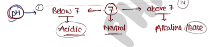
- pH: Potential of hydrogen, It is the measure of the concn of hydrogen ions ($H^+$) in body.

Mechanism : The kidney uses several mechanism to regulate acid-base balance.
- Excreting excess ions : The kidneys excrete excess ion in the urine (in PCT), which helps to remove excess acid from the body.
- Reabsorbing bicarbonate ions : The kidney reabsorb bicarbonate ion ($HCO_3^-$) from the filtrate back into bloodstream, which helps to maintain stable pH. (PCT reabsorb bicarbonate ions and secretes ions).
- Regulating electrolyte levels : The kidney regulates electrolyte levels, such as sodium, potassium and calcium, which help to maintain acid-base balance.
- Producing Ammonia : The kidney produce ammonia ($NH_3$), which helps to buffer excess ions in urine.
- Adjusting pH : Kidney has also ability to maintain the pH of urine to maintain acid-base balance.
ROLE OF RAAS in KIDNEY
RAAS Renin Angiotensin Aldosterone System
- It play an important role in regulating blood pressure, fluid balance and electrolyte balance in body.

- It starts, when liver release Angiotensinogen and kidney releases Renin.
- It mainly increases the fluid retention, reabsorption and Blood pressure.