Human Anatomy And Physiology 2 - Unit 2
Syllabus
Digestive system
Anatomy of GI Tract with special reference to anatomy and functions of stomach, ( Acid production in the stomach, regulation of acid production through parasympathetic nervous system, pepsin role in protein digestion) small intestine and large intestine, anatomy and functions of salivary glands, pancreas and liver, movements of GIT, digestion and absorption of nutrients and disorders of GIT.
Energetics
Formation and role of ATP, Creatinine Phosphate and BMR.
Scroll to Download
HAP-II
UNIT-II
DIGESTIVE SYSTEM
IMPORTANT QUESTION
Describe in detailed note on Anatomy and Physiology of Digestive system. OR Discuss various types organs and functions of Digestive system with well labelled diagram + Physiology of Digestion + Hormones regulating digestive activities.
Write a note on:-
i. Role of pepsin in digestion of protein
ii. structure of liver and role of bile juice in digestion.
iii. functions of Salivary gland and small intestineDiscuss Acid production in stomach and Regulation of acid production through PNS.
Write a note on mechanism of ATP formation and BMR.
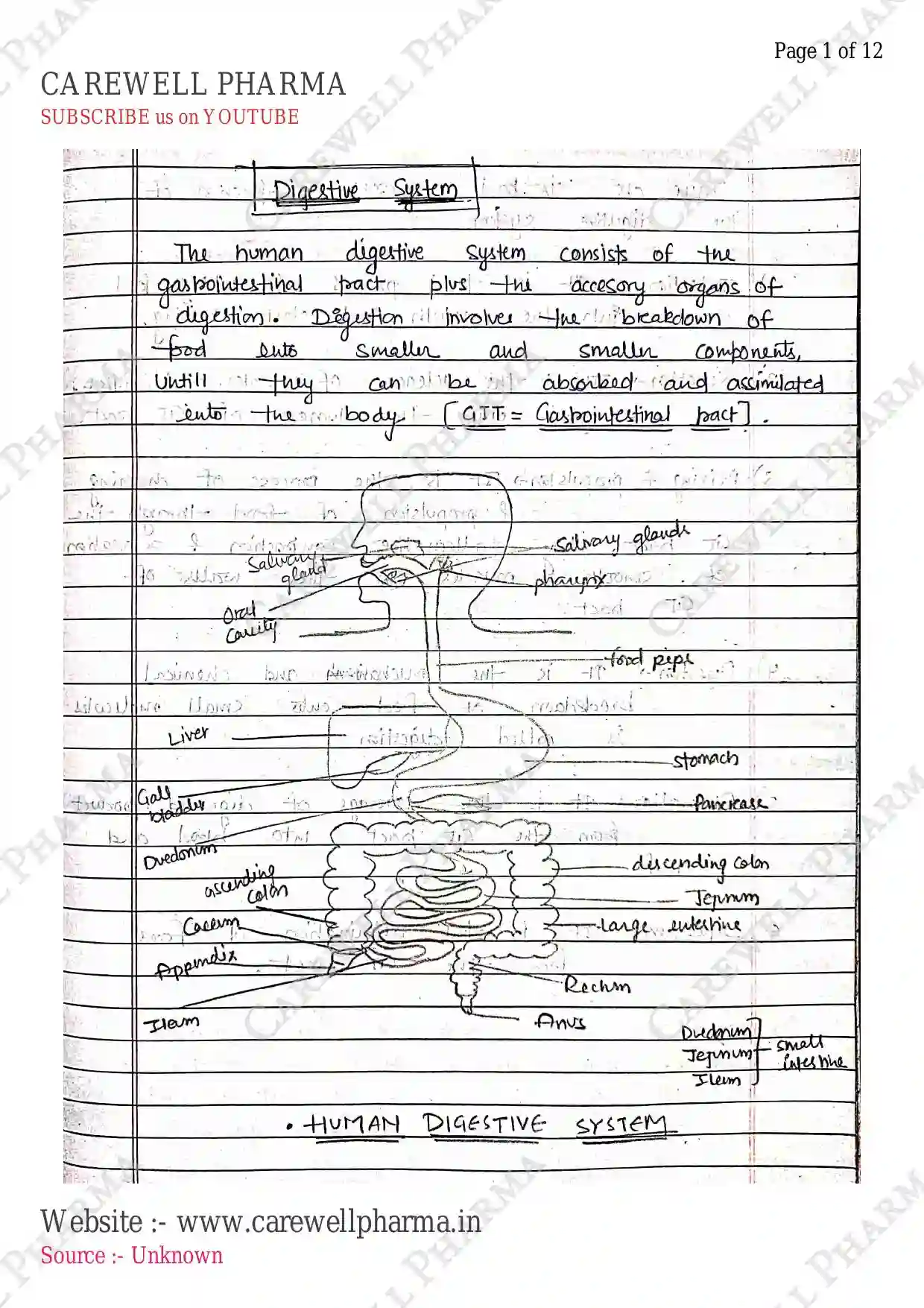
DIGESTIVE SYSTEM
- Digestive system is a network of organs and tissue that work together to perform Digestion.
- Digestion is a complex process that involves the breakdown and absorption of nutrients from ingested food.
- This system consists of Gastrointestinal (GI) tract, Accesory organs and the digestive processes.

- The GI tract is a long, hollow tubes extends from mouth to Anus, including, esophagus, stomach, small and large intestine.
- Accesory Organs: Liver, Gallbladder, Pancreas and salivary glands.
The Digestive system has mainly four functions:-
- Ingestion: food enters the mouth.
- Digestion: It involves mechanical (chewing) & chemical digestion (enzymes break down nutrients).
- Absorption: Nutrients are absorbed into the blood.
- Elimination: Waste is eliminated through feces.
ANATOMY AND PHYSIOLOGY
- Digestive system includes organs from mouth to Anus.
- It consists of:-
I) Mouth/oral Cavity
II) Pharynx
III) Oesophagus/Esophagus
IV) Stomach
V) Small intestine
VI) Large intestine
VII) Rectum / Anus
- It also consists of Accesory organs, that support the GI tract in digestion:
i. Salivary gland
ii. Liver
iii. Gall bladder
iv. Pancreas
Layers of GI Track
GI tract is composed of four types of layers:
- Mucosa : It is the innermost layer, composed of epithelial cells, lamina propria and muscularis mucosae. It produces mucosa (protection), contact luminal contents, absorption, secretion (e.g. gastric juice).

Submucosa : made up of loose connective tissue, contains nerves, lymphatic vessels, glands. It provide support, blood supply and nerve supply.
Muscularis : layer of smooth muscles, responsible for motility/movement and peristalsis.
Serosa/Adventitia : outermost layer, covers the GI tract, provide protection and support.
I) Oral Cavity (Mouth)
- It is also known as Oral cavity/Buccal cavity.
- It is the begining of GI tract / Alimentary canal.
- It consists of:-
- Teeth: Break down food into smaller pieces (mechanical digestion / Chewing).
- Tongue: mixes food with saliva and helps in swallowing of food.
- Salivary gland: produce saliva which contains enzymes (amylase & lipase) helps to breakdown Carbohydrates and fats.

- Other parts:
- Space b/w gums & cheek is Vestibule.
- Roof of mouth is palate.
- The main functions of mouth is:-
- Ingestion of food (entry of food).
- Mechanical breakdown of food by teeth.
- formation of bolus (food + Saliva) moisten of food.
- mixing and swallowing of food (help of tongue).
II) Pharynx (throat)
- It is a muscular funnel shaped tube that passes the bolus (food) from mouth to esophagus.
- It is common pathway for both respiratory and digestive system.

- It consists of:-
- Nasopharynx (connect to Nose)
- Oropharynx (connect to mouth)
- Laryngopharynx (connect to larynx)
- At the time of eating/ingestion, it opens the food pipe / esophagus and closes the trachea to avoid the entry of food in trachea (Epiglottis).
III) Oesophagus/Esophagus
- It is also known as food pipe. It is a long muscular tube (approx. long) which main work is to passes the food from pharynx to the stomach at gastro-esophageal junctions.
- It contains sphincters at its upper and lower ends to prevent the backflow of food.
IV) Stomach
- J-shaped hollow, bag-like structure.
- It lie b/w esophagus & small intestine.

- Approx. long, wide (it vary in every individuals).
- It is divided into four regions:-
- Cardia: It connect esophagus to the stomach. It contain sphincter which prevent backflow of food or acid.
- Fundus: It is dome-shaped upper part of stomach, which contains various cells such as parietal cells produce , chief cells produce Pepsin, Renin, Lipase.
- Body/corpus: It is the central region of stomach, which act as main site of action for digestion of food.
- Pylorus / Pyloric Antrum: It is the lower part of stomach, which connects the stomach to the small intestine through duodenum. It transport chyme to the duodenum. It also contain G-cells which secretes Gastrin hormones.
- It is also made up of four layers:- serosa (outermost), muscle layer (muscularis), submucosa & mucosa (in the form of large folds called rugae).
- Mucosa is responsible for gastric secretion.
Physiology/functions of Stomach
- Mechanical breakdown of food & it used for temporary storage of food.
- chemical digestion of food by digestive enzymes (pepsinogen, gastric amylase and gastric lipase) + ($HCl$) acid.
- formation of chyme (food + Acid + digestive enzymes).
- It performs secretion of Gastric juice, including:- which helps in digestion:
- Mucus
- Pepsinogen (pepsin)
- Gastric amylase and lipase.
- It performs:- Peristalsis movement of food.
- denaturation of proteins (release amino acid).
- breakdown of carbohydrates and fat by bile juice.
V) Small Intestine
It is a long, thin tube which perform final digestion of chyme (partially digested food).

- It is about long.
- It is responsible for the absorption of nutrients.
- It contains three parts:-
- Duodenum
- Jejunum
- Ileum
- The walls of small intestine contains finger-like projection called villi, which increases the surface area for absorption.
VI) Large intestine
- The large intestine, also known as colon is a wider tube that absorb water and electrolytes (ions & vitamines).

- It is about in length & in diameter.
- It contain four parts: Ascending, Transverse, descending colon & Sigmoid colon.
- It also includes Caecum (pouch), appendix (vermiform).
- After absorption, it eliminates the waste/faeces through Anus.
VII) Rectum
- It is the end part of large intestine, and about in length.
- It stores faeces temporary, as it get filled, the walls of rectum expands which activates stretch receptor, which stimulates the nervous system which helps in emptying the bowel.
VIII) Anus (Anal Cavity)
- It is the external opening of the rectum. It helps in the removal of faeces from body.
- Contains two sphincters, internally sphincter made up of smooth muscle and is Involuntary. but external sphincter is voluntary.
ACCESORY ORGANS
- There are four organs, which are outside the GI tract but helps in the digestion.
1) Salivary Gland

- It is located in the mouth and throat and produce Saliva (a clear liquid) that helps in digestion, health. (water ($99%$), enzyme, mucus, electrolytes).
It contain:-
- Parotid glands: It is the largest salivary gland, location just in front of the ear. It secretes saliva through cheek.
- Submandibular glands: located under the jawbone, these glands produce most of the saliva.
- Sublingual glands: located under the tongue, these glands produce a small amount of saliva.
- Minor salivary glands: small glands located throughout the mouth & throat.
functions:
- It moistens the food to make it easier to swallow.
- Saliva helps to break down food.
- It contains enzymes like amylase and lipase that breakdown carbohydrates and fats.
- It also contains antibodies that help to fight off infections.
2) Liver

- The liver is a vital organ located in the upper right side of the abdominal cavity.
- It is the largest internal organ in the human body.
- Size: Approx. weight and long.
- It is divided into two major lobes (left and right) and two minor lobes (caudate and quadrate).
- It is reddish-brown color in human.
- It also consists of:-
- Blood supply: Hepatic artery ($O_2$), Hepatic vein ($CO_2$).
- Liver Lobules: functional units composed of hepatocytes, sinosoids and bile canaliculi.
- Liver cells (Hepatocytes): responsible for liver functions surrounded by sinosoids (blood vessels).
functions of Liver:
- Detoxification: removing toxins/waste products from the blood.
- Metabolism: Breaking down nutrients & producing energy.
- Production of bile: bile produced by hepatocytes, stored in gallbladder and released into small intestine to perform digestion.
- It also stores glycogen to regulate blood sugar levels.
Bile Ducts:
- It carry bile (secretion of liver) which are formed by the union of biliary canaliculi.
- It unites right and left lobes to form hepatic duct which unites with pancreatic duct in duodenum (at papilla) & released bile into small intestine.
3) Gall bladder
It is a pear-shaped, hollow structure located under the liver that play a role in digestion.
- The main function of gall bladder is to store the bile, which is produced by liver.
- Then it concentrated the bile by removing excess water and electrolytes and released into in small intestine for digestion & absorption.
Role of Bile Juice in digestion:
- It play a major role in the breakdown and absorption of fats. It also play a various role:-
- Emulsification - break fats into smaller droplets.
- Activates lipase (break triglycerides into fatty acids & glycerol).
- Absorption of fat-soluble vitamines (Vit. A, D, E, K).
- Also have anti-microbial properties (eliminates pathogens/microbes).
- stimulation of gut motility.
4) Pancreas

- It is a vital organ located behind the stomach that play a important role in digestion & glucose regulation.
- It is located behind the stomach.
- Approx. ($15 \text{ cm}$) long, & wide.
- It consist of Head, Body & tail.
- It act as both exocrine and endocrine.
- Exocrine secretion: It secretes pancreatic juice that helps in digestion through pancreatic duct in S.I.
- Digestive enzymes (Amylase, Lipase, trypsin) to breaks carbohydrates, fats and proteins.
- Bicarbonate (to regulate acid balance) & water.
PHYSIOLOGY OF DIGESTION
Digestion : It is the complete process of breaking down food into smaller molecules that can be absorbed into the body through blood.
These are the main functions of digestive system:-

HORMONAL REGULATION
- Gastrin: stimulates gastric juice and acid.
- Secretin: stimulates pancreatic juice and bile.
- Cholecystokinin (CCK): stimulates pancreatic juice, bile and gastric emptying.
- Insulin: regulates glucose absorption & blood sugar levels.
- Glucagon: regulates glucose release.
ACID PRODUCTION IN THE STOMACH
- It is a complex process, which involves the production of (Acid) in the stomach from the parietal cell.
- HCl is a strong acid, which is essential to maintain a low pH (1-3) in stomach. to maintain digestion of protein or killing ingested bacteria.
- GI tract contains four layers.
In this, mucus membrane contains gastric gland.

- The production of is regulated by the PNS and hormones such as Gastrin and histamine.
- It involves several steps:-


HCl formed inside lumen of stomach and helps in digestion.
REGULATION OF ACID PRODUCTION BY PNS
Parasympathetic Nervous system play a crucial role in regulating acid production:-
- Cephalic phase - It starts before the food enters the stomach,

Vagus nerve stimulation - The vagus nerve, a major PNS nerve, release acetylcholine, which stimulates parietal cells to produce .
Gastrin release - The PNS stimulates G-cell to release Gastrin, which stimulates parietal cells to produce HCl.
Histamine release - The PNS stimulates Enterochromaffin-like cells to release histamine, which stimulates to release HCl.
- PNS also regulates acid production in response to food intake, stress and other factors.
- During eating, the PNS stimulates acid production to facilitate protein digestion.
- During stress, the PNS stimulates acid production, which can lead to increased risk of peptic ulcers.
- Somatostatin, secretin and prostaglandins inhibit acid production.
- Dysregulation of acid production can lead to disorders such as gastroesophageal reflux disease (GERD) and Zollinger-Ellison syndrome.
ROLE OF PEPSIN IN DIGESTION
- Pepsin is a digestive enzyme that breaks down protein into smaller particle of peptides and amino acids.
- It helps in digestion of food.
Pepsin formation : Chief cells in stomach produce & secrete an inactive enzyme precursor.

Protein denaturation : Activated pepsin is then secreted into the stomach lumen, where it can digest proteins.
HCl denatures proteins, unwinding their complex structures and making them accessible to pepsin.

Other functions:
- Activates other digestive enzymes.
- Pepsin work optimally at pH of 2-3, which is maintained by in stomach.
- also helps in the digestion of other foods, maintaining gastric health.
ENERGETICS
Syllabus : Formation and Role of ATP, Creatine Phosphate and BMR.
Energetics: It is the study of energy and its distribution in chemical, physical or biological activities.
Energy: It is act as a currency for our body, which is used to perform function in our body.
- It is measured and expressed in joules (unit of work) or kilocalories (unit of heat).
- In a human body, of energy per day is produced through metabolic reactions.
- eg. of Carbohydrates provides .
- of protein provides .
- of fat provides .

ATP (Adenosine Triphosphate)
- It is the primary energy currency of the cell, which store and release energy for the functioning of cells.
- It is made up of:-
- Adenine (nitrogenous base)
- Ribose (five - carbon sugar)
- Three phosphate groups (triphosphate).
FORMATION OF ATP
- It is formed through a process called cellular respiration which involves the breakdown of glucose and other organic molecules to produce energy.
- There are main three stages of cellular respiration.
- Glycolysis
- Krebs cycle/Citric Acid Cycle
- Oxidative phosphorylation / ETC
- Glycolysis : It involves carbohydrates metabolism, occurs in cell cytosol. Glucose is converted into pyruvate, producing a amount of ATP and NADH.
- Energy Input: 2 ATP investment
- Energy output: 4 ATP earn & 2 NADH earn
- Net yield: . .
- Kreb's cycle : Pyruvate is converted into Acetyl-CoA, which enters the citric acid cycle, producing more ATP, NADH, .
- Energy input: 2 ATP
- Energy output: , ,
- Total:
- Profit = from 2 mol of Acetyl CoA.
- Oxidative Phosphorylation : The electrons from NADH and are passed through a series of protein complexes in the mitochondrial inner membrane, producing a proton gradient. ATP synthase uses this gradient to produce ATP.
- Total energy yield: approx.
- Also produced from lipid, AA metabolism.
ROLE OF ATP
It play a various role:-
- Energy Currency: Primary energy source for cellular processes.
- Muscle Contraction: ATP is necessary for muscle contraction & relaxation.
- Protein synthesis: It is also required for protein synthesis & folding.
- Membrane transport: ATP is needed for active transport of molecules across cell membranes.
- Cell signalling: ATP is involved in cell signalling pathway.
- DNA synthesis: ATP is necessary for DNA replication & repair.
- Cell division: ATP is required for cell division and cytokines.
FORMATION AND ROLE OF CREATINE PHOSPHATE
- Creatine Phosphate is a high energy compound stored in muscle cells, serving as a rapid energy source to refill ATP stores during intense muscle activity.
- Also known as Phosphocreatine (PCr).
- It is storage form of energy in the muscle.

Role
- Energy buffering: act as high energy reserve, refill ATP store during intense muscle activity.
- ATP regeneration: donate its phosphate group to ADP, to reform ATP.
- helps in muscle contraction, especially for high-intensity activities.
BMR (Basal Metabolic Rate)
- The rate at which metabolic reactions use energy.
- It is the number of calories required in a body to functions at rest.
- It is the minimum energy requirement to maintain the basic body functions, when body is in rest condition.
- It includes:-
- working of heart and other organs
- conduction of nerve impulse
- reabsorption by renal tubules
- ions transport across membranes
- GI motility
Normal Value
- Adult man
- Adult woman
- Normal to variations.
Measurement of BMR By Harris-Benedict method
- For men .
- For women .
Mifflin St. Jeor equation:- BMR (calories)
- For men
- For women
FACTOR AFFECTING BMR
- Age: BMR decreases with Age.
- Sex: men generally have a higher BMR than woman.
- Weight: BMR increases with body weight.
- Height: Taller individuals have generally high BMR than short.
- Genetic: Genetic variations can influence BMR.
- Physical activity: BMR increases with regular exercise.
- Starvation: During starvation, a decrease in BMR upto has been reported.
- Environmental factors: Climate, altitude, and diet can also affect BMR.
SIGNIFICANCE/IMPORTANCE OF BMR
- BMR is important to calculate the caloric requirement of an individuals, which helps in planning of diets, weight loss/weight gain strategy.
- BMR changes can indicate underlying health issues.
- BMR is below normal in starvation, under nutrition, disease.
- BMR is above normal in fever, diabetes patients, leukemia and polycythemia.