Human Anatomy And Physiology 2 - Unit 1
Syllabus
Nervous system
Organization of nervous system, neuron, neuroglia, classification and properties of nerve fibre, electrophysiology, action potential, nerve impulse, receptors, synapse, neurotransmitters. Central nervous system: Meninges, ventricles of brain and cerebrospinal fluid.structure and functions of brain (cerebrum, brain stem, cerebellum), spinal cord (gross structure, functions of afferent and efferent nerve tracts,reflex activity)
Scroll to Download
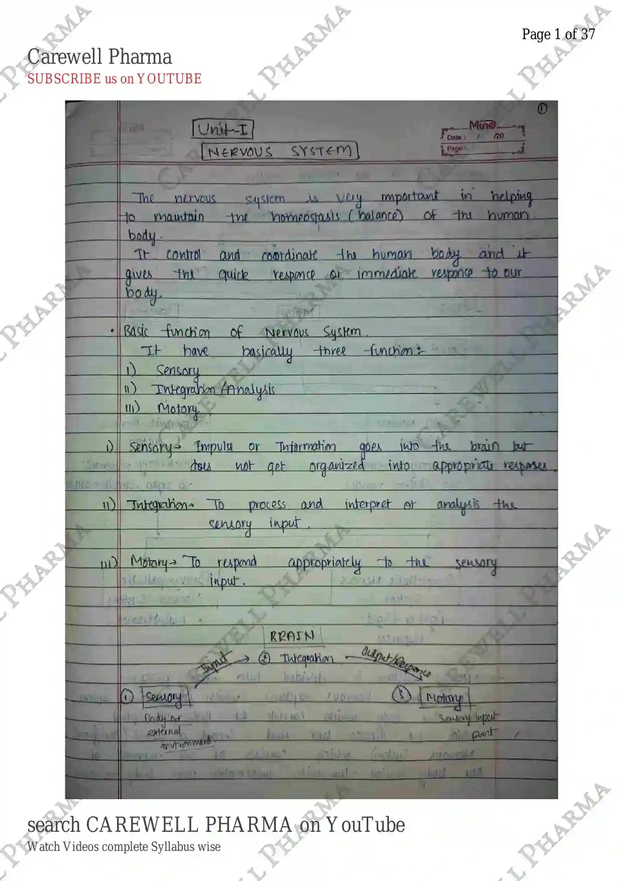
NERVOUS SYSTEM
Unit-I
The nervous system is very important in helping to maintain the homeostasis (balance) of the human body.
It control and coordinate the human body and it gives the quick responce of immediate responce to our body.
Basic function of Nervous System
It have basically three function:
I) Sensory
II) Integration/Analysis
III) Motory
I) Sensory Impulse or Information goes into the brain but basically not get organized into appropriate responce.
II) Integration To process and interpret or analysis the sensory input.
III) Motory To respond appropriately to the sensory input.

Organization of the Nervous system

Nervous system is divided into two parts:
- CNS (Central nervous system) which is main system of our body which consists of Brain & spinal cord. which we discuss later. And
- Second is PNS (Peripheral Nervous System) which consists of all nerves of our body which transmit information from body to brain.
Sensory Nervous System In which Impulse or information goes to brain from peripheral nervous system in our body.
Motor Nervous System It is the response of our CNS to peripheral nervous system.
Somatic Nervous System In this system, voluntary movement happened, ex. hand movement, walking etc. which we can do by him or her self.
Autonomic Nervous System These are the involuntary responce of our body which we can't control on it. Breathing, digestion, Heart rate etc. maintain by this Nervous System.
Sympathetic Nervous System In this system, when our body is in emergency sitution. That are when we are in any abnormal sitution or fight or flight sitution it helps. High blood pressure, decreass digestion rate and increass heart rate etc.
Parasympathetic Nervous System When our body come back from Sympathetic or any stress or any flight or fight sitution it maintain it and back to its normal condition. It Increase digestion rate and normal heart rate etc.
Nervous System made up with Nervous tissue

Neurons (Nerve cells)
It is the basic element of the nervous system. transmit information to other nerve cells, muscle or gland cells. Most neurons have a cell body, an axon, and dendrites. The cell body contains the nucleus and cytoplasm.
These are Amitotic in nature in which cell division not happened. Centrioles is absent so chromosome not separate.

Parts of Neurons:
I) Cell body It is also called Soma. It is main processing centre of the cell. It consist nucleus and organelles but absent of centrioles. and it has high metabolic rate and produce grey matter and Nissl bodies.
II) Dendrites These are thin branching extensions of the cell body that conduct nerve impulse towards the cell body. It also have extension of dendrites, dendritic spines.
III) Axon It is the extension of the cell body. single branch which conduct nerve impulses away from the cell body.
It is also called nerve fiber.

- Axon start from the point which is called Axon hillock and Action potential is also generated in Axon hillock.
- And plasma membrane of Axon is known as axolemma, which is covered by Schwann cells.
- Schwann cell is just outside of axolemma and it gives myelin sheath to the Axon.
- And Myelin sheath it is make by neurilemma (fatty material of schwann cell) And Myelin sheath is outside the Axon, provide safety. insulate the axon.
Node of Ranvier It is boundary of Schwann cells where myelin sheath is absent. And impulses jump node to node.
Axon terminal At the end, Axon ends with synaptic knob which is also known as axon terminal. Axon terminal contain vessicles inside it, and contain neurotransmitter inside it which transfer information from one neuron to another.
- Tracts Bundle of nerve fibres in CNS.
- Nerves Bundle of nerve fibres in PNS (Peripheral N.S.).
Classification of Neurons

On Basis of function:
Sensory Neuron These are receptor neurons which carry impulse from sensory organs to CNS. There are nerve which send impulse from sensory organs to CNS.
Motor Neuron These are those neurons which carry impulse from CNS to effector organs like muscles or glands.
Interneuron These are those neurons which connects sensory and motor neurons. These associated both neuron so it is also called Association neuron.
On basis of structure:
I) Multipolar Neuron These are those neurons in which cell body having multiple processes.

II) Bipolar Neuron These are those neuron in which cell body having only two processes

III) Unipolar Neuron These are those neuron in which cell body having only one processes or extension only and then it divide into two. (Contains: dendrites, peripheral process, cell body, central process, Axon).

Neuroglial cells
- It is also known as Neuroglia cells or glial cells.
Neuro + Glial $\rightarrow$ Nerve + Glue
So These are those cells which provide structural framework and metabolic support to the neurone.
- These cells fill spaces, provide structural framework produce neyelin and carry on phagocytosis.
- And neuroglial cells perform mechanical and metabolic Support roles for neurons to ensure their survival.

In CNS
- Astrocytes: It get their name due to the fact that they look somewhat like a star.
- It usually sits between a neuron cell body and a blood capillary. It prevent Toxins and poison from entering into neurons out of the brain.
- Regulate the flow of ions, sugars, oxygen & carbon dioxide of neurons.
- Ependymal cells: Usually cube-shaped.
- It form the lining of the brain's central cavities (ventricles) and Spinal cord central canal. Protects the brain and spinal cord mechanically and immunologically. The clear Liquid that fills these cavities is cerebrospinal fluid.
- It have cilia to help circulate CSF.
- Microglial cells: It is smallest of the neuroglial cells.
- It is able to migrate around the CNS.
- It support neuron by phagocytizing bacteria cells and dead cell debris.
- Oligodendrocytes: occur in rows along nerve fibers.
- They provide insulating layers of myelin around axon within the brain and spinal cord.
- Myelin helps to speed up the transmission of electrical signals in neurons.
In PNS
- Schwann cells: The PNS contain neuroglial cells as well known as Schwann cells
- It form a myelin sheath (covering) around axons.
- Basically they perform the same roll as the oligodendrocytes found in the CNS.
- Satellite cells: These are small glia that surround neurons sensory ganglia in the ANS.
- PNS Satellite glia are very sensitive to injury and may exacerbate (खराब करना / तेज करना) pathological pain.
- It gives protection to the Neuron cells.

Electrophysiology
Neuron is a modified cell and contain cell body Which contain all simple cell organelles and nucleus. So, it contain cell body in which flow of ions takes place.
It includes measurements of the electrical activity of neurons & action potential activity it is all about the flow of ions in biological tissue.

Cell body of neurons: Every cell contain plasma membrane and There are ions present at inside and outside the plasma membrane (Neuronal membrane).
- ion (sodium ion) have high concentration at outside the cell so it is called extracellular ion.
- ion (Potassium) have high concentration at inside the cell so it is known as intracellular ion.
- Also some proteins are more inside the cells.
At Resting state (When nerve impulse is not transmitted from neurons) ion moves inside the cells and ion moves outside the cells. Because both follow law of diffusion (higher con. to lower). But neuronal membrane is not permeable for ion so it does not move inside the cell.
- But ions moves outside the cells. So when concentration inside the cell thin (-) ve charge produce at inside the plasma membrane and (+) ve charge at outside the plasma membrane.
- So, due to presence of charge at membrane So it is Polarized.
Due to change concentration of ion at inside and outside the cells it develop potential difference.
At resting state (Resting potential).
Sodium Potassium pump manage that difference and then Na^+$/$K^+ into its equal concentration back.
- Potential Change: RMP (Resting Membrane Potential = $-70mV$)
Thresold Potential
When any kind of Stimulus (pressure, temp etc.) detected by neurons OR when neurotransmitter send stimulus to neurons it changes the Resting membrane potential to less potential and it is called depolarisation.

If Stimulus change Resting membrane potential ($-70mV$) to then it is called Threshold potential.
If Stimulus achieved threshold potential then it generate Action potential and further it generate Nerve impulse.
Action Potential:
When Stimulus achieved threshold potential It open channel. So, ions enters inside the cell. then due to more ion inside the cell there are change of charge at plasma membrane takes place and (+) ve produce at inside the plasma membrane and (-)ve at outside the cells. And it is called Depolarisation.
- At same time channel also open and outflux of ion takes place. So ion moves outside the cell and again charges changes and (-)ve at inside and (+)ve at outside it is called Repolarisation.


- The cycle of depolarisation and repolarisation is called Action potential.
- These Action potential works 1000 times in one second.
Nerve Impulse
It is a signal that transmitted along a nerve fibres. In generally, it is Information or stimulus that is transfer through the neurons. these are the electrical signal which is generated or produced or transmitted through the Action potential.
- It is transmitted through Axon by propagation of action potential.

- Nerve Impulse is transmitted like this through action potential.
- And propogation of action potential is nerve impulse.
Receptors (Neurotransmitter Receptors) (Neuroreceptor)
Receptors are those binding sites which are made up with proteins. Neurotransmitter receptors are present in post synaptic membrane.
Types of Receptors:
I) Ionotropic receptors (Ligand-gated ion channels) (LGICs).

- Neurotransmitter binding sites and Ion channel both are associated.
- When neurotransmitter attached to their site it open or close ion channel according to neurotransmitter nature.
II) Metabotropic receptors (G-Protein coupled receptors) (GPCRs)

- Neurotransmitter binding sites and Ion channel both are in different or separated membrane.
- And Receptors attached with G-Protein.
- When neurotransmitter attached to their receptor's sites it will directly open or close ion channel or by indirectly (through secondary messenger).
Synapse
It is a Junction of two neuron in CNS.

Ganglia - And also a Junction of two neuron but in PNS
- Synapse is made up b/w one neuron to another Neuron or, Neurons to effector organs.
Types of Synapse :
- Electrical Synapse (fast transmission)
- Chemical Synapse
I) Electrical Synapse :
In an electrical synapse, the axon of one neuron (presynaptic neuron) is connected to the dendrites of another neuron (post synaptic neuron) by gap junction. The gap junction is composed of sets of channel. Each channel is made up of six protein subunit. known as a Connexon.
And two connexon, one in presynaptic membrane and second in postsynaptic membrane make up a gap junction. And Gap junction allow small molecules and ions to pass freely from the cytosol of one cells to another. So, it allow to Action potential, Nerve impulse to pass across the gap junction.

II) Chemical Synapse :
In chemical synapse, nerve impulse is transmitted through chemical from one neuron to another.
- In between one neuron to another neuron there are some distance approx. 20-30 nm, that is called Synaptic cleft.
- At the end of the neuron, in axon terminal there are synaptic vessicles are present and Neurotransmitter is present in these vesicles.
- So, When nerve impulse (Action potential) reached at pre synaptic neuron it rapture the vessicles at terminal.
- Then these vesicles release neurotransmitter in synaptic cleft, And it is received by receptors. which is present on post-synaptic membrane.
- So, nerve impulse transmitted through the chemical. so, it is called chemical synapse.

Neurotransmitter
Neurotransmitters are chemical messengers that transmit signals from a neuron to a target cell across a synapse.
- Target cell may be a neuron or some other kind of cell like a muscle or gland cell.
- It is packaged into synaptic vesicle presynaptic side of a synapse.
Types of neurotransmitters :
- Inhibitory: GABA, Serotonin
- Excitatory: Glutamate, Aspartate
- Both: Acetylcholine, Norepinephrine, Dopamine
Functions:
- Adrenaline (fight or flight) - It is produced in stressful situation, increase heart rate and blood flow,
- leading to physical boost and heightened awareness.
- Noradrenaline (Norepinephrine) (concentration) - it affect attention and responding actions in the Brain.
- Contracts blood vessels increasing blood flow.
- Dopamine (pleasure) - feeling of pleasure, also addiction movement and motivation.
- People repeat behaviours that lead to dopamine release.
- Serotonin (mood) - Contributes to well-beings and happiness.
- Helps sleep cycle and digestive system regulation.
- Affected by exercise and Light exposure.
- GABA (Calming) - Calms firing nerves in the central nervous system.
- High levels improve focus.
- Low levels cause anxiety.
- Also contributes to motor control and vision.
- Acetylcholine (Learning) - Involved in thought, learning and memory. activates muscle action in the body.
- Also associated with attention and awakening.
- Glutamate (memory) - Most common neurotransmitter. Involved in learning and memory.
- It regulates development and creation of nerve contacts.
- Endorphins (Euphoria)
- Released during exercise, excitement and sex
- Producing well-being and euphoria
- Reducing pain.
CNS (Central Nervous System)
The central nervous system (CNS) is the part of nervous system consisting primarily the brain and spinal cord.
The central nervous system (CNS) is responsible for integrating sensory information and respond accordingly.
Meninges
It is covering of the brain. Nervous tissue is not a sturdy tissue. Even moderate pressure can kill nerve cells, so it is surrounded by three fluid containing membrane called the meninges. It provide protection & support to the CNS.
- And then meninges is surrounded by bone.
- The spinal meninges form a tube like covering around the spinal cord and line the bony vertebral foramen of the vertebrae that surrounded the cord.
It contain three layers:
I) Dura mater (Outermost layer)
II) Arachnoid mater (middle layer)
III) Pia mater (Innermost layer)
- Outer layer Dura mater which is the tough that Lines the vertebral canal which is just below Skull.

- Pia meter the innermost layer which is connect on the Cerebrum.
- Arachnoid mater is present b/w the Dura mater and Pia mater.
- There are Subdural space present b/w the Dura mater And Arachnoid mater And Subarachnoid space present b/w the Arachnoid mater and Pia mater. it have a spidery like structure and it contain fluid filled with CSF (Cerebrospinal fluid).
- So, Meninges are present b/w the bony structure and the nervous tissue to provide protection to the structure.
Ventricles of brain and cerebro spinal fluid (CSF)
- Most part of the brain is hollow and from inside there are various cavities called ventricles. And production and circulation of CSF (Cerebrospinal fluid) takes place in ventricles. The cavities are filled up with CSF.
- So, Lining of these ventricles are made up with ependymal cells which is glial cells.
- Ependymal cell and Capillaries which is together form a choroid plexus responsible for the production of CSF.
- There are four ventricles in brain. The largest ventricles are those which are present in the cerebrum there are two of them which are placed in the form of lateral ventricles as one in each hemisphere.
- The third ventricle is a tubular space lying in the middle of diencephalon which is connected on one side with a lateral ventricle through a passage called interventricular foramen or foramen of munro while on the other end it continues into the fourth ventricle through cerebral aqueduct. fourth ventricle which is present b/w brainstem and cerebellum.
CSF (Cerebrospinal fluid)
is a colorless & clear Liquid which occupies the cavities of the brain & spinal cord which is secreted by choroid plexus.
- Composition: It contain water, glucose, creatine, electrolytes such as chlorides of calcium, sodium and potassium.
- It is approximately 180 ml in adults. It is present in brain and spinal cord.
- Functions: It is meant for acting as a cushion against shocks or jerks and also a medium for the movement of nutrients, metabolic waste and exchange of respiratory gases. It provides mechanical and chemical protection and circulation of nutrients. It also maintain Homeostatic balance.
Circulation of CSF : It is formed in the choroid plexus of each lateral ventricle and then flow into the third ventricle through foramen of monro. Third ventricle also produce CSF which mix with CSF whose come from lateral ventricle then all flow into fourth ventricle through cerebral aqueduct. And the choroid plexus of the fourth ventricle contribute more fluid.
- Then CSF enters the subarachnoid space through three opening in the roof of the fourth ventricle (a median aperture and the paired lateral apertures). CSF then circulate in the central canal of the spinal cord & in the subarachnoid space around the surface of the brain and spinal cord.
- CSF is gradually reabsorbed into the blood through arachnoid villi (through venous) then it goes into heart and lungs and then reabsorbed by blood.

Brain
Brain is largest organ of soft nervous system (tissue) contained in the skull of vertebrates, functioning as the coordinating center of the body.
- It is one of the largest organs in the body and coordinates most body actiities. thought, memory, judgement and emotions.
- Each part of the brain is responsible for controlling specific functions, such as temperature, regulation and breathing.
- The brain is contained in skull and weight is 1300-1400gm. Made up of 100 billion neuron, & each neuron is surrounded by about 10 glial cells.
- Neurons cannot multiply & many neurons are lost everyday in life but glial cell can multiply throughout the life.
- Brain is also covered by meninges.

Cerebrum
It is the largest section of the brain and located in the upper portion of the brain.
- The outermost layer of cerebrum is made up of grey mater and this is 2-4 mm thick and it is called "Cerebral Cortex". It contain millions of neurons.
- During embryonic development, the brain size increases rapidly, the gray mater of cortex enlarges much faster than deeper white mater so as result cortical region rolls and folds upon itself.
- The folds are called "gyri" or "convolutions".
- the deepest grooves b/w the folds are called "fissures".
- the shallows grooves b/w folds are called "sulci".
- The most prominent fissure, the longitudinal fissure, separates the Cerebrum into right and left halves called "cerebral hemisphere". Each hemisphere has 4 lobes.
- Both Right and left hemisphere are connected by a bridge of nerve fibre that relay (share) information b/w two hemisphere called "corpus callosum".
- Left hemisphere controls right side of the body.
- Right hemisphere controls left side of the body.


Lobes of cerebrum:
- Frontal lobe : most anterior portion of the cerebrum (under forehead) "central sulcus" separate frontal & parietal lobe.
- Control motor function, personality, and speech.
- Like centre of reasoning, planning some parts of speech, movement, emotions, problem solving.
- Also called "motor cortex".
- Parietal lobe : The most superior portion of the cerebrum (top of head), it receive and interpret nerve impulses from sensory receptors.
- It receive sensory input from the skin (touch, pressure, temperature and pain).
- Also called "sensory cortex".
- Occipital lobe : The most posterior portion of the cerebrum (back of the head).
- It receive input from the eyes. control visions.
- Also called as "visual cortex".
- Temporal lobe : The left and right lateral portion of the cerebrum (on the side of your head above ears).
- It controls hearing and smell.
- Also called "Auditory cortex".
Functions of cerebrum:
- motor functions like control of voluntary movement.
- Sensory functions like perception of pain, temperature, touch, pressure, hearing, taste and smell.
- Control of intelligence, speech, memory and learning etc.
Cerebellum
It is the second largest portion of the brain.
- It is located beneath the posterior part of the cerebrum.
- A deep groove known as "transverse fissure" separate cerebrum to cerebellum.
- Aids in coordinating voluntary body movements and maintaining balance and equilibrium.
Structure :
- The external surface called "cerebellar cortex" look like a butterfly and lateral wings or lobes called cerebellar hemisphere that is interconnected by a narrow portion called vermis.
- It contain 10% weight of entire brain and 50% of neurons.
I) Anterior lobe
II) Posterior lobe - both regulate skeleton muscle movement.
III) Flocculonodular lobe - on the inferior surface maintain equilibrium & balance.
Cerebellar peduncles :
3 paired attach the cerebellum to brain stem.
- Superior cerebellar peduncle: is a paired structure that connects the cerebellum to the midbrain.
- Middle cerebellar peduncle: connect cerebellum to pons.
- Inferior cerebellar peduncles: is a thick rope-like strand that occupies part of the posterior district of the upper half of the medulla oblongata.
Functions of cerebellum :
- Coordinate contractions of skeletal muscles.
- Regulate posture & balance.
- May play a role in learning from experiences & language processing.
Brain Stem
consists of Midbrain, Pons and medulla oblongata.
- Superiorly Continuous with Diencephalon superiorly and interiorly continuous with spinal cord.
I) Mid brain : it is located below the cerebral cortex and about 2.5cm long.
- It Act as pathway for impulses to be conducted b/w the brain and spinal cord.
- Screening of information before it reach high brain structure.
- It control reflex movement of the body and hearing reflexes.
II) Pons: It means bridge - connects the cerebellum to the rest of the brain. And it is located above medulla oblongata and below midbrain.
- It Controls sleep as well as the rate and pattern of breathing.
III) Medulla Oblongata: It is posterior part of the brain. Controls automatic actions of our body such as: Breathing, Swallowing, Heart rate, Blood circulation.
Diencephalon
Posterior part of the forebrain that connects the midbrain with the cerebral hemisphere.
I) Thalamus - It is located above the brain stem and b/w the cerebral cortex and mid brain.
- It carries sensory information from the body to the cerebrum and the Limbic system.
II) Hypothalamus - lies under the thalamus.
- It connects the nervous system with the endocrine system via pituitary glands.
III) Pituitary gland - It is a pea-shaped structure and It is very important in growth and reproduction.
- And it also maintain other harmone gland.
IV) Epithalamus & Pineal gland - Epithalamus is a small region superior & posterior to the thalamus and it consist of pineal gland.
- Pineal gland worked as biological watch for our body which control or maintain our rhythm.
The Cranial nerves
The 12 pairs of cranial nerves named because they pass through bones of cranium & arise from the brain inside the cranial cavity. These nerves may be sensory, motor or mixed (both).
| Number | Name | Composition | Function |
|---|---|---|---|
| I | Olfactory | Sensory | olfaction (smell). |
| II | Optic | Sensory | Sight or Vision. |
| III | Oculomotor | Mixed | moves eyes, pupil. |
| IV | Trochlear | Mixed | moves eye. |
| V | Trigeminal | Mixed | face sensation. |
| VI | Abducens | Mixed | moves eye. |
| VII | Facial | Mixed | moves face, salivate. |
| VIII | Vestibulocochlear | Sensory | hearing, balance. |
| IX | Glossopharyngeal | Mixed | taste, swallow. |
| X | Vagus | Mixed | heart rate, digestion. |
| XI | Accessory | Mixed | moves head. |
| XII | Hypoglossal | Mixed | moves tongue. |
Mixed: Motor and sensory both.
Spinal cord
The spinal cord is continuous with the brain and lies caudally to the brain, protected by the vertebra and spinal meninges.
- It starts from the base of the skull, starting below the foramen magnum and terminates at the first or second lumbar vertebra.
- It is a slender column of nervous tissue continues with brain and brainstem.
- Extends downward through vertebral canal protected by the spinal meninges.
- The spinal cord and spinal nerves serve two functions.
- Pathway for sensory and motor.
- Responsible for reflexes activity (functional independence).
Structure of spinal cord :
Spinal cord is situated inside the vertebral canal. Length of Spinal cord (16-18 inches) and diameter - 2cm.
- Spinal cord starts from medulla oblongata and protected by spinal meninges. Typical adult length ranges b/w 42-45 cm.
- It runs to the lower lumbar or to the lower of 1st and 2nd lumbar vertebrae (in new born till 3-4 Lumbar grows yr stops but vertebral canal grows till adulthood).
- So, The spinal cord is shorter than the vertebral canal that houses it.

External anatomy of spinal cord
Two enlargements in spinal cord at:
- Cervical (C4 to T1) - nerves to upper limbs.
- Lumbar (T9 - T12) - nerves to lower limbs.
Conus medullaris - It is the tapered inferior end (conical) of the spinal cord below lumbar enlargement b/w the 1st and 2nd lumbar vertebrae, which marks the official "end" of the spinal cord.
Cauda equina (horse tail) - They are inferior to conus medullaris, which forms collection of nerve roots (lumbar, sacral, coccyx-groups of axon) that project and leave inferiorly from the spinal cord.
Filum terminale - They are structures within the cauda equina consists of thin strands of pia mater, which helps to anchor the conus medullaris to the coccyx.
The spinal cord is associated with 31 pairs of spinal nerves, which connects the CNS to receptors and Effectors (muscles and glands).
Roughly cylindrical elongated lower part of CNS.
Slightly flattened both posteriorly and anteriorly.
External surface has two longitudinal depressions:
The posterior (dorsal) median sulcus
The anterior (ventral) median fissure
Internal anatomy
- It is composed of white mater in which H shaped (or butterfly shaped) grey mater present in between white mater.
- Because of present of grey mater in between white mater, White mater divided into three region or column.
- Posterior white column (Back side)
- Anterior white column (front side)
- lateral white column (side ways)

- Grey mater present in H shaped in posterior and anterior side.
- It contain posterior and anterior horn.
- Central canal present in spinal cord in which CSF present.
- Roots - Bundle of axon connect nerve to spinal nuclei.
- posterior root or posterior horn (sensory part enter).
- Anterior root or anterior horn (motor part leave, exit).
- Tracts - Ascending tracts or Afferent parts conduct sensory impulses to the brain And Descending tracts or efferent parts conduct motor impulses from the brain to motor neuron reaching muscles and glands.
- Afferent nerves - carry impulses / information like touch, pain, vibration, pressure etc from peripheral receptors to higher centres in the brain through ascending tracts of spinal cord.
- Efferent nerves - Carry command from higher centres of CNS to effector organs like muscles etc... for execution of an action through the descending tracts of spinal cord.
Functions of spinal cord
- The pathway for nerve impulses Conduct to and from brain and brainstem.
- Center for spinal reflex.
Reflex activity
It is also known as reflex arc.
- It is rapid predictable and unplanned response to a stimuli.
- Due to any type of stimuli which our body receive our body give rapid responce to that stimuli rapid, and unplanned and it is known as reflexes.
- Reflexes are automatic, subconscious response to stimuli within or outside the body.
It contain five components:
I) Receptor - also known as sensory receptors, which receive stimuli. Then it transmits that nerve impulse.
II) Sensory neuron - pass on stimuli to the spinal cord.
III) Inter neurons - serves as a processing center: conduct nerve impulse from sensory neurons to motor neuron. also known as association neuron.
IV) Motor neuron - transmit that nerve impulse from the spinal cord to an effector organ.
V) Effector - responds to stimulation by the motor neuron and produces the reflex or behavioral action. It may be a muscle or gland.

- It helps to maintain homeostasis (e.g. heart rate, breathing, blood pressure etc.)
- also helps to maintain automatic action (e.g. swallowing, sneezing, coughing etc.)
- also helps to maintain body balance and body posture.