Biochemistry - Unit 4
Syllabus
Nucleic acid metabolism and genetic information transfer
Biosynthesis of purine and pyrimidine nucleotides Catabolism of purine nucleotides and Hyperuricemia and Gout disease Organization of mammalian genome Structure of DNA and RNA and their functions DNA replication (semi conservative model) Transcription or RNA synthesis Genetic code, Translation or Protein synthesis and inhibitors
Scroll to Download
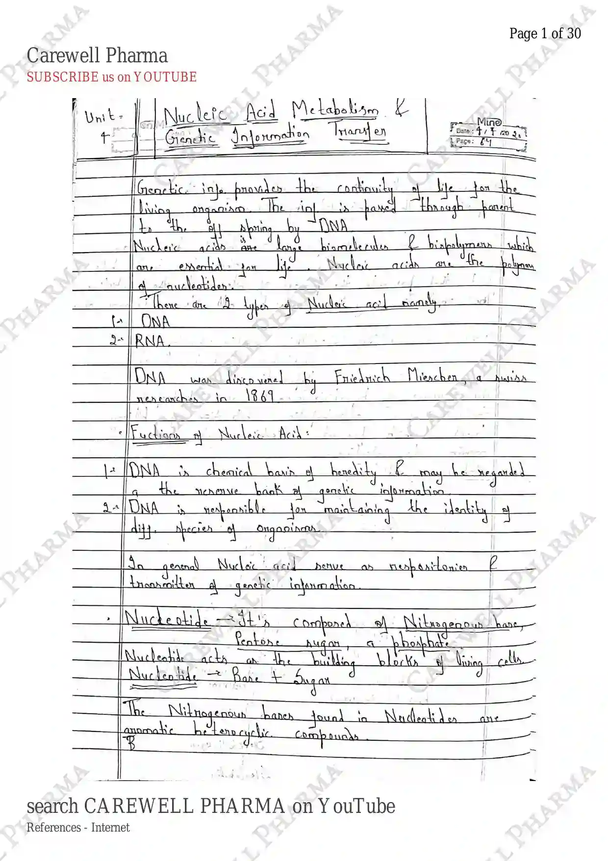
NUCLEIC ACID METABOLISM
UNIT-4
SYLLABUS
Nucleic acid Metabolism and Genetic information transfer: Biosynthesis of purine and pyrimidine nucleotides, Catabolism of purine nucleotides and Hyperuricemia and Gout disease organisation of mammalian genome, structure of DNA and RNA and their functions, DNA replication (semi conservative model), Transcription or RNA synthesis, Genetic code Translation or Protein synthesis and inhibitors.
INTRODUCTION

- Nucleic acid are those molecules, which is made up by the polymerisation of nucleotide.
- Nucleotides are the building block of nucleic acid, which consist of Pentose sugar, Phosphate group and a Nitrogenous base.
- Nucleic acids are those naturally occuring molecules which are inside nucleus (nuclei) of cells and contain genetic information in the form of DNA and RNA.
- DNA was discovered by friedrich miescher in 1869.
- Nucleic Acid metabolism involves the synthesis, modification and degradation of nucleic acid. This is important for maintenance, replication and expression of genetic information.
- It involve several processes like synthesis of nucleotides, DNA replication, RNA synthesis (transcription) and catabolism of nucleotides.
NUCLEOTIDE BIOSYNTHESIS
- Nucleotides are the building blocks of Nucleic acid. It is composed of three parts :-
- Pentose sugar
- Phosphate group
- Nitrogenous base
1. Pentose sugar : These are those carbohydrates which contain 5 carbon. Two types of sugar associates with Nucleic Acid.

2. Phosphate group : It is also a part of nucleotides that connects with sugar. It contain three hydroxyl group and one Oxygen linked with phosphorus. (ester bond)

3. Nitrogenous base : these are nitrogenous heterocyclic compounds containing nitrogen (N).
- Purines: these are double ringed nitrogenous compounds. (Adenine, Guanine)
- Pyrimidines: these are single ringed nitrogenous compounds. (Uracil, thymine, Cytosine)

BIOSYNTHESIS OF PURINE NUCLEOTIDES
- It can be performed by two methods:-
- De novo pathway
- Salvage pathway
- Adenine and Guanine (purine nitrogenous bases) are not synthesized as a free base, instead they synthesized in the form of purine nucleotide i.e. Adenosine-Monophosphate (AMP) (for adenine) and Guanosine-monophosphate (GMP) (for Guanine).
De Novo synthesis/pathway of purine synthesis
- In this, synthesis is starting from begining, for this firstly we have to synthesized IMP (Inosine monophosphate) a precursor.
Synthesis of Inosine Monophosphate (IMP)
- It starts from the activation of Ribose-5-Phosphate and formation of PRPP (5-Phosphoribosyl-1-pyrophosphate).
- It ends at the formation of Inosine monophosphate (IMP) which further used in the formation of AMP and GMP.
- It involves utilisation of lots of energy.

Synthesis of AMP (Adenosine Monophosphate) & GMP

Synthesis of AMP from IMP
- It involves the synthesis of AMP by forming Adenylosuccinate in the presence of enzyme synthase by utilising GTP & Aspartate.
- Then formed AMP by lyase enzyme & releasing fumarate.
Synthesis of GMP from IMP
- Frstly IMP formed XMP by enzyme IMP dehydrogenase by utilising energy .
- Wich then formed GMP by enzyme synthetase & utilising ATP and Glutamine.
Salvage Pathway of Purine synthesis
- In this pathway, free purines (occured from metabolism of N.A.) reused for the synthesis of purine nucleotides.
- It involves the recycling of the free bases.

BIOSYNTHESIS OF PYRIMIDINE NUCLEOTIDES
- It is also be prepared by de novo and salvage pathway:
De novo pathway of pyrimidine synthesis
- It involves the synthesis of pyrimidine ring.
- It is synthesized in the form of pyrimidine nucleotides :-
- Cytidine Monophosphate (CMP)
- Uridine Monophosphate (UMP), and
- Thymidine Monophosphate (TMP).

- , Glutamine and ATP form Carbamoyl phosphate by using enzyme CP synthase-II (for urea synthesis).
- Now, Carbamoyl phosphate condense with Aspartic acid to form Carbamoyl aspartate (CA) by using enzyme Aspartate transcarbamoylase.
- Then this further formed UDP (Uridine Diphosphate) by several reactions and several enzymes from step 3 to step 7.
- Now, this UDP further formed UTP by using ATP which further synthesized CTP (Cytidine Triphosphate) by using enzyme synthase.
- On other side, UDP formed dUDP (deoxyuridine diphosphate) by using ribonucleotide reductase and utilising NADPH.
- Then, dUDP formed dUMP and finally formed dTMP by using thymidylate synthase.
Salvage pathway for pyrimidine
- In this, pyrimidine nucleotides are formed by free pyrimidine bases.

DEGRADATION OF NUCLEOTIDES
- It involves the catabolism of purines and pyrimidines
1. Catabolism of Purines

2. Catabolism of Pyrimidine Nucleotides

HYPERURICEMIA
- It is a condition of excess Uric acid in the blood.
- In this condition, uric acid passes through liver and reaches the bloodstream.
- Normally, uric acid is excreted in the form of urine.
Causes:
- It is mainly caused due to increased levels of uric acids.
- Primary hyperuricemia It is produced in large amount due to purines and excretion of uric acid does not occurs.
- Secondary hyperuricemia It is uric acid due to another disease/condition.
GOUT
- It is a condition occurs due to deposition of monosodium urate (uric acid) accumulates within the tissue in excessive amounts, leading to inflammation and pain in joints (painful joints) i.e. most common inflammatory arthritis.
- It is associated with elevated levels of uric acid in the blood (hyperuricemia) and this uric acid are produced by the catabolism of purines.
Causes:
- It is occurs due to overproduction of uric acid and it occurs :-
- genetic factor (mutation in genes involves purine metabolism)
- increased purine intake (Red meat, sea food, high fructose)
- cell turnover (condition such as leukemia, psoriasis etc)
- due to underexcretion of uric acid
- renal impairment
- genetic factor
Crystal formation:
- Monosodium urate deposites mainly in the joints in the form of crystals.
- Monosodium urate crystals are formed when uric acid levels exceed its solubility limits (around 6-8mg/dl), the blood become supersaturated.
- This leads to the precipitation of monosodium urate crystals especially in color part of the body such as joints.

STRUCTURE & FUNCTIONS OF DNA & RNA
- Nucleic acids are of two types:
- DNA
- RNA
- Nucleic acids are biopolymers of nucleotides components.
DNA
It refers to Deoxy Ribo Nucleic Acid
It consists of Nucleotides contains
- Deoxyribose sugar with double stranded sugar phosphate backbone.
- Nitrogenous bases such as (Purines: Adenine, Guanine) and (Pyrimidines: Cytosine, Thymine).
- phosphate group.
Deoxyribose sugars: These Carbohydrates contain 5 carbons.

- Phosphate Groups: linked point.

- Nitrogenous Base: Adenine, Guanine, Cytosine, Thymine.


Double helix:
It consisting of two strands that coil around each other to form a double helix.
- These strands are antiparallel, means they run in opposite direction i.e. 5' to 3' and 3' to 5'.
- The backbone of each strand is made up of alternating phosphate & deoxyribose sugar molecules. (sugar phosphate).
STRUCTURE
- In this, Nitrogenous bases are paired with H-bond
- Nucleotides are attached with phosphodiester bonds.

FUNCTIONS OF DNA
- Genetic information It is a genetic material which contain/store genetic information (eg. color of skin, eye, height, Intelligence) in the form of long sequences of bases. It transfer information from parents to their daughter cells.
- Replication DNA can replicate itself & passed DNA during cell divison.
- Transcription DNA serve as a template for the synthesis of RNA.
- Translation Information on all cellular protein synthesis is carried by DNA. This segment is known as gene.
RNA
- It refers to Ribo Nucleic Acid
- It contains ribose pentose sugar and are single stranded.
- It contains nitrogenous bases Adenine, Guanine, Cytosine, Uracil.
- It contains phosphate group.
Types / Components:
- RNA in a cell is present in amount 10 times more than that of DNA, because RNA performs various cellular functions.
- It exists in several forms, with unique functions:-
- r-RNA (Ribosomal RNA) It is the most abundant RNA, which participates in protein synthesis to provide binding site on the ribosome for t-RNA and m-RNA.
- t-RNA (Transfer RNA) It brings amino acids to the ribosomes during protein synthesis.
- m-RNA (messenger RNA) It is least abundant (5-10%) and it carries genetic information from DNA to ribosome, where proteins are synthesized.
- snRNA (small Nuclear RNA) involves in RNA splicing.
- miRNA (Micro RNA) and siRNA (small interfering RNA) involved in gene regulation.

Single Standerd
- It is typically single stranded but can form complex three dimensional structures through intramolecular base pairing.
- In this, Nitrogenous bases are paired with H-bond: , , , .
- These are attached with phosphodiester bond.

FUNCTIONS OF RNA
- Protein Synthesis RNA is synthesized from DNA in the nucleus. The resulting mRNA carries the genetic code from the DNA to the ribosomes. (Transcription). Then this transcription follow translation and synthesized proteins with the help of tRNA.
- Gene Regulation RNA molecules like miRNA and siRNA regulates gene expression by degradation of mRNA.
- mRNA (messenger RNA) transfer genetic information from nucleus to the cytoplasm.
- tRNA (transfer RNA) works as carrier of amino acids to the site of protein synthesis.
- rRNA (ribosomal RNA) is required for the formation of ribosomes.
DEFINITIONS
DNA Deoxyribo Nucleic Acid is a double stranded molecules that stores genetic information and transfer it to new cell during cell division. It contain deoxyribose sugar, phosphate group and nitrogenous bases ($A=T$, $C\equiv G$). It also serves as template for RNA synthesis.
RNA Ribo Nucleic Acid is a single stranded molecules synthesized from DNA and play a major role in synthesis of proteins, regulating gene expression and catalyzing biological reactions. It contain ribose sugar, phosphate group and nitrogenous bases ($A=U$, $C\equiv G$). In this, thymine is replaced by uracil.
DNA REPLICATION
- It is a process by which double stranded DNA molecules is copied to produce two identical DNA molecules.
- It is self-replicating structure and replicates semi-conservatively.
Semiconservative Model:
- Acc. to this, DNA get separated its double strands into two single strands during replication then each strand work as a template for the synthesis of new DNA strand.
- So, it forms a new DNA with one old and one new strand.

Process of DNA replication
- It involves various steps and also various enzymes.
Steps:
- firstly, synthesis of DNA initiates at the site "origin of replication" where replication originated. In eukaryotes there are multiple 'Ori'.
- Now, specific protein dnaA binds at 'Ori' and initiate replication & form replication bubble at site.
- Then, the catalyze enzyme Helicase unzip the DNA helix by breaking H-bond b/w base pairs and forms Replication fork. obtain energy from ATP.
- Now, SSBP (single strand binding proteins) attached these single strands to prevent intercoiling of DNA.
- Now, the main enzyme DNA polymerase starts synthesis by adding nucleotides to parent strand, but it works on some conditions.
- It required a primer to start (RNA primer, a short fragment of RNA synthesized by the enzyme DNA primase) which further replaced.
- It works unidirectional i.e. 5' to 3' know as leading strands and forms continuous replication and other is discontinuous replication.
- On lagging strands, a short Okazaki fragments are formed. After synthesis, exonuclease remove them, DNA polymerase formed new & DNA ligase joined & sealed them to form continuous strands.
- During separation, Topoisomerase enzymes relieves the stress/supercoiling generated by unwinding of DNA.

RNA SYNTHESIS (Transcription)
- It is the first step of gene expression that involves the formation of RNA molecules from DNA.
- Gene expression is the process of using genetic information stored in DNA for biological function i.e. transcription and translation.
- DNA RNA Protein
- Transcription process is highly selective (RNA molecules are synthesized from only some DNA regions).
- RNA polymerase is the main enzyme to perform transcription.
STEPS
It occurs in three steps:
- Initiation
- Elongation
- Termination
- Initiation: firstly RNA polymerase attaches to the DNA molecules and moves along to recognise promoter sequence (starting site of transcription).
- Now DNA double helix unwinds and only one strands works as a template for RNA synthesis i.e. 3' to 5', because RNA synthesized at 5' to 3'.

- Elongation: in this, Ribonucleotides are added to the template strand to growth the mRNA but proper adding base pairs. (, , , $C \equiv G$).

- Termination: Now, After synthesis of RNA encounter terminator region which stops synthesis.
- Now, RNA polymerase releases & pre-mRNA not ready to leave nucleus.

- Maturation of RNA: In this, A Amino acids sequences i.e. methylated guonine cap and Polyadernylation tail are added to mRNA to protect in from degradation.
- At last, splicing occurs in this non-coding sequences are removed by spliceasome excision.
- Lastly, a mature mRNA is obtained which further used in the synthesis of proteins.
PROTEIN SYNTHESIS / TRANSLATION
- Translation is the 2nd step in gene expression.
- Translation is the process of synthesis of proteins from mRNA.
Components:
- mRNA: It contains genetic information in the form of codons.
- tRNA: It transfers amino acids to the ribosomes, acc to sequences.

STEPS
It occurs in three steps:
- Initiation
- Elongation
- Termination
- Initiation: firstly mRNA is attached to the smaller subunit of ribosome.
- Now t-RNA containing Amino acids (methionine) are attached to these mRNA and starts reading the codons.
- when t-RNA encounter AUG codons, it immediatly starts the synthesis of protein by binding larger subunits to smaller subunits and form complete ribosomes.
- Elongation: Now, methionine (t-RNA) attached at P-site of ribosomes, further tRNA matches the next codon and starts the formation of peptide linkage of amino acids.
- Now, t-RNA (methionine) goes to E-site by releasing amino acids (met) to P-site, and next t-RNA moves to A-site, then to P-site and A-site vacate for next amino acids (t-RNA).
- This cycle repeats and P-site holds the polypeptide chains. & new amino acids joined by peptide linkage.
- Termination: It is the last step, in this when t-RNA encounter any one codons (UAG) (UAA) (UGA) i.e. terminating factors, it release polypeptides chain of proteins & stop translation.
- After this, some process like amino acid deletion and protein folding occurs which then forms proteins.

Inhibitors of Protein synthesis
- These are those molecules that can interfere with the various stages of protein synthesis.
- These inhibitors are important tools in research & medicines, particularly as antibiotics for bacterial infections and as agents in cancer treatments.
- e.g., Streptomycin, tetracycline, chloramphenicol, erythromycin, ricin.
Genetic Code
- It is the information contained by the m-RNA and this information is used for the synthesis of proteins.
- It exists as codons, these are a group of 3 adjacent bases specifying the amino acids of proteins.
- It is made up of three nucleotide (triplet) base sequences in m-RNA acting as code words for amino acids in proteins.
- It composed of four nucleotide base
- purine Bases Adenine (A) and Guanine (G)
- pyrimidine Bases Cytosine (C) and Uracil (U).

Structure of tRNA
- complementary binding of codon (of m-RNA) and Anticodons (of t-RNA)