Human Anatomy And Physiology 1 - Unit 5
Syllabus
Cardiovascular system
Heart - anatomy of heart, blood circulation, blood vessels, structure and functions of artery, vein and capillaries, elements of conduction system of heart and heart beat, its regulation by autonomic nervous system, cardiac output, cardiac cycle. Regulation of blood pressure, pulse, electrocardiogram and disorders of heart.
Scroll to Download
CARDIOVASCULAR SYSTEM
UNIT 5
SYLLABUS
Heart - Anatomy of heart, Blood circulation, Blood vessels, structure and functions of artery, vein and capillaries, elements of conduction system of Heart and Heart beat, its regulation by Autonomic Nervous system, Cardiac output, cardiac cycle. Regulation of Blood pressure, pulse, Electrocardiogram (ECG), disorders of Heart.
Definitions
CARDIOVASCULAR SYSTEM Cardio (Heart) + Vascular (Blood vessels)
- Cardiovascular system composed of heart, blood vessels and blood.
- It is responsible for the transportation of Oxygen [$O_2$], nutrients to all over the body. (cells and tissue).
- It is also responsible for the transportation of and waste products from body (tissues) to lungs and other excretory organs (kidney etc).
- It is also known as circulatory system.
- It also responsible for the transportation of water, electrolytes and hormones by using blood.
- Blood is pumped through the heart with the help of blood vessels. (blood is flowing through these vessels).
HEART
- It is roughly cone shaped hollow muscular organs. It is about 10 cm long and it is about the owner's fist.
- The average weight of heart is about 250gm in adult females and 300gm in adult males.
- Location It is lie in the thoracic cavity in the space b/w the lungs [Two thirt part of heart is on left side], and behind the sternum.
LAYERS OF HEART
Heart wall is composed of three layers.
- Pericardium
- Myocardium
- Endocardium
1. Pericardium It is the outermost layer of heart that covers the heart and blood vessels Joint with it.
The main function of this layer is to protects the heart.
It is of two types:-
- Fibrous pericardium - tough, inelastic, made up with dense irregular connective tissue. It prevents overstretching, provide protection & holds heart.
- Serous pericardium - thin, delicate membrane, secretes pericardial fluids (serous fluids) which filled in cavity and protects the heart from friction and erosion.
2. Myocardium - It is the middle layer of heart and made up with Cardiac muscle tissue.
- It makes up the bulk of heart and responsible for the pumping action of heart.
3. Endocardium - It is the innermost layer of heart and made up with thin layer of epithelial tissue. It provides the smooth lining for the chambers of heart and covers the valves of heart.
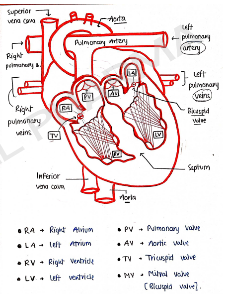
CHAMBERS OF HEART
- Heart is made up with (four) chambers, which helps in the circulation of blood.
- Right and left sides of the heart act as two separate pump.
- Left sides of heart have much larger work load than the right side, also the wall of left side is thick as compared to right.
- Right side/ventricle pump blood only to lungs, but left ventricle pump to all over the body.
It consists of :-
- Right Atrium
- Right ventricles
- Left atrium
- Left ventricles
i) Right atrium [R.A.] - small thin walled chamber
- receive deoxygenated blood from vena cava and further pumped into right ventricles.
ii) Left Atrium [L.A.] - small then right atrium but thick.
- receive oxygenated blood from lungs through pulmonary veins and pumped into left ventricle.
iii) Right Ventricle [R.V.] - larger part of heart
- receive deoxygenated blood from right atrium and pumped into lungs for exchange of gases.
iv) Left ventricle [L.V.] - below left atrium, workload
- receive oxygenated blood from left atrium and pumped into all over the body through aorta.
VALVES OF HEART
These are located within the chambers of heart and control the direction of blood flow.
Tricuspid valve - It is right atrioventricular valve situated b/w right atrium and right ventricle.
- passes blood from atrium to ventricle & prevent backflow.
Mitral valve - It is left atrioventricular valve, passes blood from left atrium to left ventricle and also prevents backflow. Also known as Bicuspid valve.
Pulmonary valve - It is semilunar valve, situated b/w right ventricle and pulmonary artery. prevent backflow.
Aortic valve - It is semilunar valve, situated b/w aorta and left ventricle. prevent backflow of blood into left ventricle.
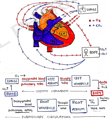
BLOOD CIRCULATION
The main function of heart is to circulate the blood to all over the body.
- During circulation, oxygen and nutrients deliver to the tissue and , waste material carry away from the tissues.
It is of two types -
- Systemic circulation: blood to all over body.
- Pulmonary circulation: blood from body to lungs.
Process
Firstly oxygenated blood [$O_2$] pumped into left atrium from lungs through pulmonary veins.
Left atrium pumped blood into left ventricle through Bicuspid/Mitral valve.
Then, this blood pumped to all body through aorta. In body, and nutrients delivered and $CO_2$/waste carried.
Then this deoxygenated [$CO_2$] blood pumped into Right Atrium through inferior and superior vena cava.
Right atrium pumped blood into right ventricle through Tricuspid valve (prevent backflow).
Then this blood pumped into lungs through pulmonary artery, release & carry and further starts circulation.
CONDUCTION SYSTEM OF HEART
- Heart is made up with myocardiac muscle (tissue) also known as Cardia cells.
- These cells have ability to generate electrical impulse itself known as Auto rythmicity and are responsible for pumping action of heart.
- They itself generate action potential/electric impulse [current to pump heart].
It consists of four major element
- SA Node
- AV Node
- Bundle of His
- Purkinje fibres
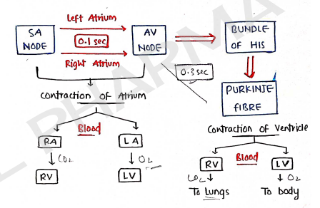
i) SA Node - It is Sino Atrial Node. It is the natural pacemaker of the heart.
- It generate action potential (electric impulse) responsible for the pumping action of heart.
- It located in the wall of right atrium near the Superior vena cava.
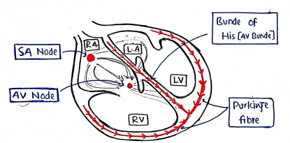
ii) AV Node - It is Atrioventricular Node.
- It is the second pacemaker of the heart.
- It located in the bottom of right atrium.
- It collects impulse from SA node and transfer to bundle of His.
iii) Bundle of His - Also known as AV bundle.
- It located b/w the atria & ventricular.
- It receives impulse from AV node and transfer to purkinje fibre.
iv) Purkinje fibre -
- It is located at the end of AV bundle at the base of heart.
- These are network of small bundle of conducting fibres and get impulse from Bundle of His.
- It is responsible for the contraction of ventricles.
HEART BEAT
- It is the rhythmic contraction and relaxation of the heart.
- Average heart beats 70-72 per min
Regulation By Autonomic Nervous system
- Sympathetic Nervous system Increase heartbeat by secreting adrenaline hormones. ($\uparrow$ depolarisation in SA node).
- Parasympathetic Nervous system decrease heart beat by secreting acetylcholine (Ach). [through vagus nerves].
CARDIAC CYCLE
- It is a complete cycle of event in the heart, from the begining of one heart beat to the begining of the next heart beat.
- It consist of contraction and relaxation of atria (auricles) and ventricles, resulting in one heart beat.
- Each cardiac cycle completed in 0.8 seconds and in three stages (average 72 bpm).
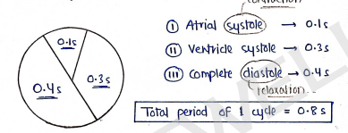
1. Atrial Systole (0.1 sec)
- It is the contraction of the heart muscle (myocardia) of the left and right atrium.
- This is marked by stimulation of the SA Node.
- In this, blood pumped from the atria into the ventricles through bicuspid and tricuspid valve.
2. Ventricle systole (0.3 sec)
- It is the contraction of the muscle of the left and right ventricle.
- It is stimulated by AV node stimulation. In this, bicuspid and tricuspid valves close and produce first heart sound i.e. lubb.
- Blood pumped into body through aorta and into lungs through pulmonary artery.
3. Complete diastole (0.4 sec)
- After contraction of ventricles, there is complete cardiac diastol, a period of 0.4 sec, when atria and ventricles are relaxed.
Total period of 1 cycle = 0.8s
CARDIAC OUTPUT
- It is defined as it is the amount of blood flowing from the heart (from left ventricle into aorta per min.) or in one heartbeat.
- where, stroke volume = volume of blood pumped by heart/heartbeat.
BLOOD VESSELS
- These are also the main part of cardiovascular system that forms a closed structure/circuit of tubules, in which blood flow from the heart to all body then bring it back to heart.
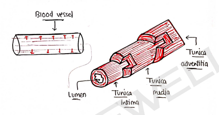
It is composed of three layers:-
Tunica adventitia: It is the outermost layer and composed of collagen and loose connective tissue.
Tunica media: It is the thickest and middle layer. It is composed of smooth muscles, elastic fibres and connective tissue.
Tunica intima: It is the innermost layer, and composed of simple squamous epithelium. It formed a hollow structure lumen in which blood flow.
Types: It is mainly of three types:-
- Artery
- Veins
- Capillaries
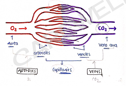
i) Artery
- These are those blood vessels that carry and flow oxygenated blood to all over the body. [arteries]... blood.
- Examples:
- Aorta: It is the largest artery which carry blood directly from heart.
- Arteriols: these are the smallest arteries which have small diameter.
- Coronary arteries: those arteries through which heart muscle get oxygenated blood.
ii) Veins
- These are those blood vessels that carry and flow deoxygenated blood from all over body to get back to heart & lungs.
- Examples:
- Vena Cava: It is the largest vein that pump (carry) blood into the heart. (i.e. inferior and Superior vena cava).
- Venules: these are the small veins that have small diameter.
iii) Capillaries
- Those blood vessels which have smallest diameter and connecting the arterioles to the venules.
BLOOD PRESSURE
- It is the pressure exerted by the blood to the inner wall of blood vessels.
- It mainly depends on the diameter of blood vessels.
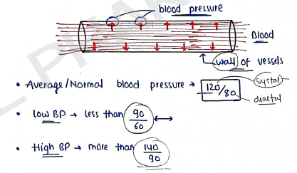
Average/Normal blood pressure 120/80 mmHg
- Low BP: less than 90/60
- High BP: more than 140/90
It is of two types:-
- Systolic blood pressure: occurs during systol of heart (i.e. ventricular contraction). Range 100-120 mmHg.
- Diastolic blood pressure: minimum pressure occurs during the diastol of heart (i.e. ventricular relaxation). Range 60-80 mmHg.
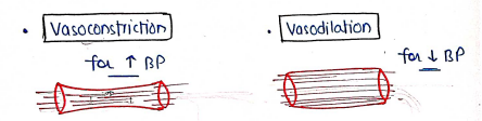
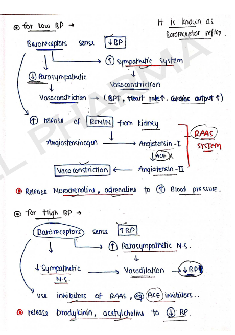
Regulation of Blood Pressure
- It is normal in change in blood pressure (i.e. Low BP and High BP).
- It depends on the diameter of blood vessels.
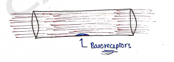
1. Baroreceptors
- These are those receptors which are located in blood vessels in Carotid sinus and in aortic arch.
- These are sensitive for pressure change which detects changes in blood pressure through the level of stretch on vascular walls.
ELECTROCARDIOGRAM (ECG)
- It is defined as it is the graphical representation of electrical activity of heart or heart beat.
- It is simply the recording of heart beat/rhythm.
- The device/machine which is used to measure heart beat is known as Electro Cardio Graph.
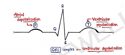
An ECG has three main components
P wave: It is the small deflection on ECG.
- It starts when the electric activity generated by SA node (pacemaker of heart).
- This electric impulse stimulates the atria and forms the P wave known as atrial depolarisation.
QRS complex: This impulse activates the main pumping chambers of heart (i.e. ventricles), and produces the large up-and-down wave in the middle, the QRS complex, known as ventricular depolarisation (contraction).
T wave: It is dome shaped upward deflection in ECG.
- It is the relaxation of ventriculars, when impulse reverses over the ventricles and travel back. known as ventricular repolarisation.
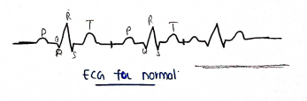
- For normal heart beat, there is regular pattern of the P wave, QRS complex and T wave.
- It takes about 0.8 sec for entire ECG cycle.
PULSE
- It is an rhythmic beat felt in an artery.
- It is an indicator of the pumping action of the heart.
- Each pulse beat means, blood is ejected into the arteries (in the system).
- It is equal to the heart beat at about (72) bpm.
- It is also defined as it is the rhythmic contraction and relaxation of the heart and its muscles.
DISORDERS OF HEART
These are problem occured in heart.
Hypertension: It is the condition of high blood pressure.
Conjestive Heart failure (CHF): It is the condition, in which heart does not pump the blood properly to meet the normal demands.
Arrhythmia: Irregular heartbeat.
- Tachycardia: fast above 100 bpm.
- Bradycardia: slow below 60 bpm.
Hypotension: It is the condition of low blood pressure.
Myocardial infarction: It is the condition of heart attack, in which heart muscle (itself heart) does not get proper blood.