Human Anatomy And Physiology 1 - Unit 1
Syllabus
Introduction to human body
Definition and scope of anatomy and physiology, levels of structural organization and body systems, basic life processes, homeostasis, basic anatomical terminology.
Cellular level of organization
Structure and functions of cell, transport across cell membrane, cell division, cell junctions. General principles of cell communication, intracellular signaling pathway activation by extracellular signal molecule, Forms of intracellular signaling: a) Contact-dependent b) Paracrine c) Synaptic d) Endocrine
Tissue level of organization
Classification of tissues, structure, location and functions of epithelial, muscular and nervous and connective tissues.
Scroll to Download
HUMAN ANATOMY AND PHYSIOLOGY - 1ST
CHAPTER - 1ST
Introduction to Human Body
- Definition and scope of anatomy and Physiology, levels of structural organisation and body system, Basic Life processes, Homeostasis, Basic anatomical terminology
Human Anatomy & Physiology (HAP)
It is defined as it is the study of structure and functions of human body.
Anatomy - It is the branch of science which deal with the study of structure of different organs of human body (body parts).
Histology: Study about tissue.
Physiology - Is the branch of science which deal with the study of functions of different organs of human body (body parts).
- Neurophysiology: study about Neurons.
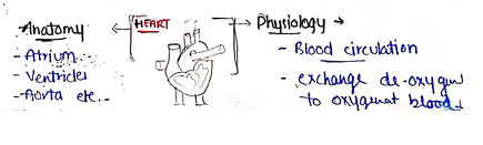
Example: HEART
- Anatomy: Atrium, Ventricles, Aorta etc..
- Physiology: Blood circulation, exchange de-oxygenated to oxygenated blood.
Scope of Anatomy & Physiology
It is about, what we can do from Anatomy & physiology.
- Study of structure and function of body parts.
- Parameters of normal health such as temp, pH, basic need etc...
- Pathology of disease.
- Complete Body Surgery techniques.
- Human evolution and development.
- Histology: study about tissue.
Levels of Structural Organisation
Human body is organised structure. Six-levels:
- Chemical level / Molecular level
- Cellular level
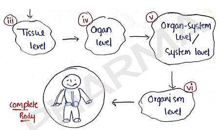
- Tissue level
- Organ level
- Organ-System level / System level
- Organism level
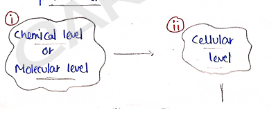
(i) Molecular/chemical level: It is the most basic level, two or more atom/molecule joined together to form cells.
- eg. Oxygen ($O$), Carbon ($C$), Hydrogen ($H$) etc.
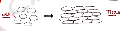
(ii) Cellular level: It is the basic structural and functional level of body. i.e. Cell. Two or more cells joined together to form tissue.
(iii) Tissue level: These are the group of cells which works together to perform a particular functions.
- eg. Nervous tissue, Epithelial tissue (skin) etc..
(iv) Organ level: In this, different-2 types of tissue combine together to form organs which do proper functioning of body.
- eg. Heart, Lungs, kidney etc.
(v) System level: In this, A group of organs combine together to form System.
- Digestive system, Respiratory system, Cardiovascular system etc.
(vi) Organism level: It is the highest level and a complete body made up with combined of all system.
- eg. Human Body.
Body Systems
A system is a group of organs, which combined together to perform proper functioning. There are total 11 systems in human body:-
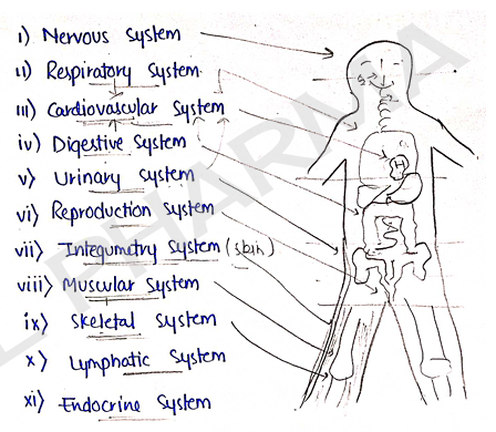
Nervous system: It coordinate all the actions of the body. It is responsible for voluntary and involuntary action and also for all signalling.
- Eg: Brain, spinal cord etc..
Respiratory System: It involves respiration ($O_2 \leftrightarrow CO_2$).
Cardiovascular System: It is responsible for the circulation of blood in body, that's why it also known as circulatory system.
- Organs: Heart, blood vessels, blood etc.
Digestive system: It is responsible for digestion of food and absorb nutrients from it.
- Organs: Mouth, stomach, intestine etc..
Urinary system: It is responsible for the filteration of blood and also removes the waste.
- Organs: kidney, Urethra, bladder etc..
Reproductive system: It is responsible for the production of offsprings.
- Organs: Testes, ovary, fallopian tube etc...
Integumentary system (skin): It is also known as exocrine system, because it contain skin which provide protection and also contain glands.
- Eg. Skin, Hair, Nails, sweat etc.
Muscular System: It is responsible for the movement of our body organs through muscles.
Skeletal system: It contain bones which maintain structure and provide protection to our body. * Skull, Ribs, femur etc...
Lymphatic System: Also known as immune system. It defends the body against pathogens that may harm the body. This system consist of network of lymphatic vessels that carry a clear fluid called lymph.
Endocrine System: A system consists of different types of hormones which helps in functioning of body.
BASIC LIFE PROCESS
Human body performs diff-2 functions for its survival and growth, so all the Living organism have some specific Life processes.
(i) Metabolism: It is the sum of all chemical process occurs in the body. It is of two types:
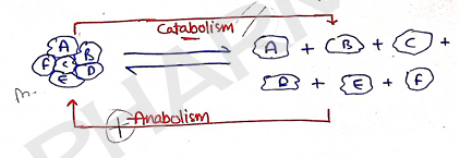
- a) Catabolism: It is the breakdown of complex chemical substance into simple compound.
- b) Anabolism: It is the building up of complex chemical substance from small components.
- (or complex molecule)
(ii) Responsiveness: It is the ability of the body to detect and respond to changes.
- eg. cold, sensitivity etc...
(iii) Movement: It includes motion of the whole body individual organs etc..
(iv) Growth: It is the development of our body and also increase in body size.
(v) Differentiation: It is the development of cell from unspecialized to a specialized state.
- eg. stem cell generate complete human body.
(vi) Reproduction: It refers to the formation of new cells and also produce new offsprings.
- eg. foetus...
(vii) Respiration: It involves the exchanges of and b/w the cell and the external environment.
(viii) Digestion: It involves the degradation of food and large molecules. It is also responsible for the absorption of nutrients into blood.
(ix) Excretion: It is the process of removal of waste products from the body.
- eg. urination etc.
HOMEOSTASIS
It is derived from two greek word:
Homeo (Same/constant) + stasis (State). It means staying the same.
Definition: It is a condition when our internal environment is constant with respect to external environment.
It is a condition that may vary but relatively remains constant.
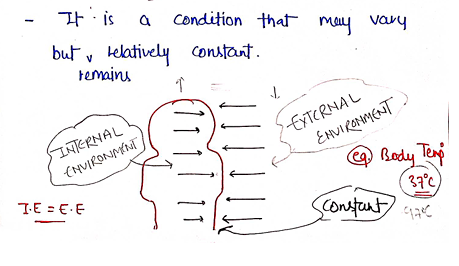
- (Balance)
- External Environment (Constant)
- eg. Body Temp
HOMEOSTASIS CONTROL MECHANISM
All the body organs coordinate with each other to maintain homeostasis. This coordination is mainly controlled by Neuroendocrine System [Nervous + Endocrine system].
It has three components:-
Receptors: It is a type of sensor, which receive/detect changes or other stimuli.
Control Centre: It receive the stimuli from receptors and analyse it.
Effectors / feedback System: If there are any change take place in internal environment, then feedback system is take back into its constant state or in homeostasis.
It is of two types:-
- Positive feedback System (+): Used to increase.
- Negative feedback system (-): Used to decrease.
(1) Positive feedback system: Used to increase. when anything is decrease in our internal environment, then it is try to back into normal situation by increasing it.
- Example: During childbirth, it stimulate the release of oxytocin which increases the Contraction of the uterus to help in childbirth.
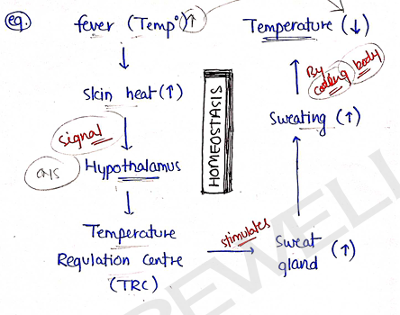
(2) Negative feedback system: Used to decrease. when anything is increase in our internal environment (body), then this system is try to back into normal condition by decreasing it.
- Example:

BASIC ANATOMICAL TERMINOLOGY
It is divided into three groups:
- Directional terms
- Sectional planes / planes of the body
- Body Cavities
1) Directional terms
- Superior (upper, cranial) upper part
- Inferior Lower part
- Anterior end front part
- Posterior end Back side
2) Sectional planes
- Sagittal plane: divides body into left and right part.
- Transverse plane: It is horizontal axis parallel to the ground and passes through the body at angle and divides body into two parts.
- Coronal plane: divides body into anterior and posterior parts.
3) Body Cavities
- Thoracic Cavity
- Abdominal & Pelvic Cavity
- Dorsal cavity
CHAPTER - 2ND
CELLULAR LEVEL OF ORGANISATION
Syllabus:
* Structure and functions of Cell
* Transport across cell membrane, Cell division, cell Junctions.
* General principles of cell communication, Intracellular signalling pathway activation by extracellular signal molecule, forms of intracellular signalling;
    * a) contact-dependent
    * b) paracrine
    * c) Synaptic
    * d) Endocrine
This is the cellular level of body which contains cells.
Cell
It is a structural, basic and functional unit of our body.
A human body consist of about or 100 trillions cells with a size and mass of about and 1 nanogram respectively.
Cells are of two types:-
- Prokaryotic cells: These are those cells which are not fully developed. They have less developed nucleus and some organelles.
- eq. Bacteria etc.
- Eukaryotic cells: These are those cells which are fully developed, they have well developed nucleus and other organelles present in cell.
- eq. plants, animals, fungi etc..
- Prokaryotic cells: These are those cells which are not fully developed. They have less developed nucleus and some organelles.
CELL
A cell is the basic living structural and functional unit of body enclosed within a membrane.
- There are about 200 different types of cells in our body.
Functions of cell:
- Movement of substance across the cell membrane.
- Cell growth and metabolism by cell division.
- Protein synthesis by transcription and translation.
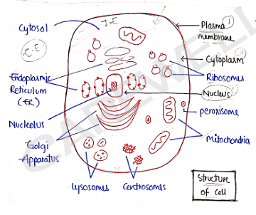
Structure of the cell: It is divided into three major parts:-
- Plasma membrane
- Cytoplasm (Cytosol + Organelles)
- Nucleus.
- Cytosol
- Endoplasmic Reticulum (ER)
- Nucleolus
- Nucleus
- Golgi Apparatus
- Lysosomes
- Centrosomes
- Peroxisome
- Mitochondria
- Ribosomes
- Cytoplasm
- Plasma Membrane
1) Plasma Membrane
Also known as Cell membrane.
- It is the outermost membrane of cell which separate out the internal environment of cell to the external environment.
- It is selectively permeable which allowing substances to pass through it.
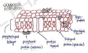
Structure: The fluid-mosaic model of plasma membrane was given by S.J. Singer and G.L. Nicholson.
It is made up with bilayer of phospholipids.
Chemical composition:
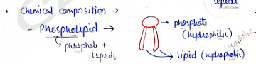
- Proteins: The lipid bilayer is embedded with proteins of various shape and sizes.
Eg. Integral, Transmembrane & Peripheral proteins
Carbohydrates: These molecules are attached with proteins (glycoprotein) and Lipids (glycolipid).

Functions:
- It provide protection from external environment.
- It give shape to the cell.
- It regulates the flow of material into and out to the cell.
- Also play a key role in communication with other cells or external environment.
2) Cytoplasm
It is the total area between the plasma membrane to the nucleus. It further divided into two parts:
- Cytosol
- Organelles
1) Cytosol: It is the fluid present inside the cells. It is transparent, viscous which contain about 75% to 90% mostly water, proteins, lipids & Carbohydrates.
2) Organelles: These are the organelles present in cytoplasm and perform specific function for cell.
- Endoplasmic Reticulum (ER)
- Ribosomes
- Mitochondria
- Golgi Apparatus / Body / Complex
- Lysosomes
- Centrosomes
- Peroxisome
Endoplasmic Reticulum (ER): It is a complex channel system enclosed within the membrane in the form of three dimensional network that consists of vesicles, flattened sacs and branched tubules.
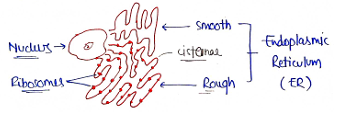
- It is of two types:-
- Rough ER: those which have ribosomes attached, helped in protein and membrane synthesis.
- Smooth ER: Ribosomes are not attached.
Functions:
- Act as a structural framework of cytoplasm.
- Exchange materials.
- SER (Smooth ER) help in synthesis of phospholipids, cholesterol and triglycerides.
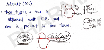
Ribosomes:
- These are tiny spheres that contain ribosomal RNA & several ribosomal proteins.
- Also known as factory of proteins, because protein synthesis is takes placed in it.
- These are made up with two subunits i.e. smaller subunits (40s) and the larger subunit (60s).
Mitochondria: It is known as the "Power House of cell", because it generate ATP.
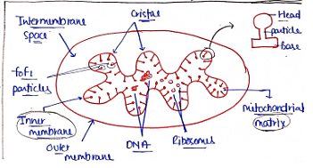
It is made up with two membranes i.e. inner and outer.
- Main function is to generate energy. Also perform cellular signalling and regulates cellular proliferation.
Golgi Apparatus / Body: It consists of four to six flattened sacs called as cisternae placed upon each other. Like a pile of plates with expanded bulges at their ends.
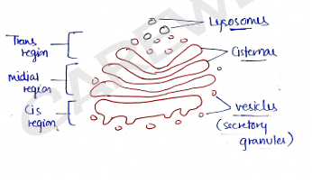
Functions: They stores proteins, modify them and also moved to the plasma membrane through secretory granules, when required.
- Also helping in excreting excess amount of water.
Lysosomes: They are secretory vesicles formed from the golgi complex.
- They worked as the digestive system of cell, which contain digestive and hydrolytic enzymes and hydrolyse large molecules, such as proteins, RNA and DNA, and Lipids.
- They vary in size ranging from 0.1-1.2 micrometer.
- They also work as defencing against microorganism.
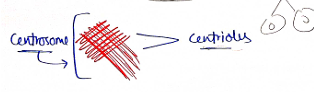
Centrosomes: It consists of a pair of centrioles and play an important role during cell division.
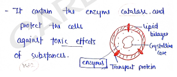
Peroxisome: It contain many oxidases enzymes that can oxidize various organic substances such as fatty acid, amino acids, uric acid etc..
- It contain the enzyme Catalase, and protect the cells against toxic effects of substances.
3) Nucleus
The nucleus is usually a spherical and oval shaped structure, which is largest in the cell.
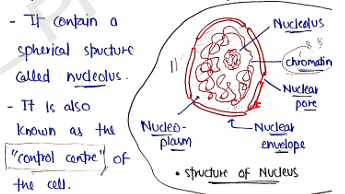
- It consist of double membrane, which separate nucleus from the Cytoplasm.
- It contain a spherical structure called nucleolus.
- It is also known as the "Control centre" of the cell.
- It contains the genetic material including aggregations of protein DNA & RNA.
- It directly involves in the reproduction and transfer all genetic information from parent cell to daughter cells.
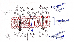
TRANSPORT ACROSS MEMBRANES
- Plasma membrane of cells are selectively permeable, which allow only some substance to cross it.
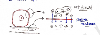
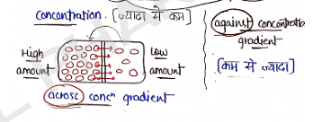
- Concentration Gradient: When any drug/substance move from high concentration to lower concentration.
Substances are transported across the cell membrane through:-
- Passive transport
- Passive diffusion
- Facilitated diffusion
- Osmosis
- Active transport
- Primary transport
- Secondary transport
- Endocytosis
i) Passive transport: In this, substance transport across the conc. gradient i.e. High to low.
- Passive diffusion: It is the transport of substance across the conc. gradient i.e. from the region of higher conc. to lower conc. without use of energy.
- eg. diffusion of lipid soluble molecules like & across the cell membrane.
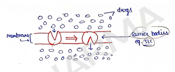
- Facilitated diffusion: In this, substance transport across the conc. gradient, but with the help of any Carrier bodies (that's why it is also called carrier mediated transport).
- It is suitable for poorly diffusible substance.
- Carrier used such as SLC (Solute carrier transporter).
- Eg. Entry of glucose into RBCs. Intestinal absorption of Vit. & .
- Osmosis: It is the movement of solvent (water) particle from high (solvent) to low conc. through semipermeable membrane. (from low solute conc. to high solute conc.).
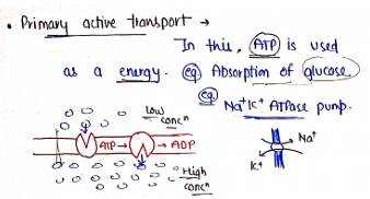
ii) Active transport: In this, substance transport against the conc. gradient with the help of energy.
In this, ATP is used.
It is of two types:-
Primary active transport: In this, ATP is used as a energy. Absorption of glucose. (Low conc. High conc.) Eg. $Na^+$-ATPase pump.
- Secondary active transport: In this, electrochemical gradient are used instead of energy. Symport (Co-transport): movement of both substance in same direction. eg $Na^+$-glucose symporter. Antiport (counter-transport): movement of molecules in opposite direction. Eg. pump.
iii) Endocytosis: In this transport, drugs of very large size molecules get transported via engulfment by cell membrane.
- eg. Cellular uptake of macromolecular like fat, starch, oil-soluble vitamins Like A, D, E, K and drug Like Insulin.
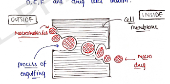
- Phagocytosis [cell eating]: absorptive uptake of solid particulates.
- Pinocytosis [cell drinking]: uptake of fluid solute.
CELL DIVISION
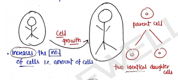
It is a process by which a parent cell divides into two daughter cells.
The genetic content (DNA) of newly formed cell is exactly same to the parent cell.
- Cell growth Increases the no. of cells i.e. amount of cells.
Cell division is of two types:-
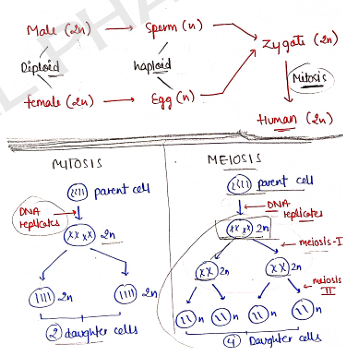
- Mitosis: responsible for growth of cells. It occurs in somatic cells. No. of chromosomes same [equational division].
- Meiosis: responsible for formation of gametes cells. occurs in reproductive cells and no. of chromosome decreased [Reductional division].
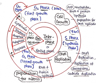
CELL CYCLE
The sequence of events by which a cell duplicates its genome, synthesis the other constituents of the cell and eventually divides into two daughter cells. It has two phases:-
- Interphase - preparation.
- M-phase - cell division.
1) Mitosis
Also known as M-phase or Mitotic phase [Equational division].
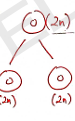
- It is occurs in somatic cell in which already existing parent cell divides into two identical daughter cells.
- In this, the no. of chromosome in daughter cell is same as the no. of chromosome in parent cell. (2n 2n)
It is occurs into two parts:-
- Karyokinesis: division of nucleus.
- Cytokinesis: division of cytoplasm.
i) Karyokinesis: It is a process by which the cell nucleus divides into two daughter nuclei.
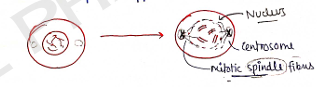
Prophase: This phase begins with initiation of condensation of the chromosomal material i.e. chromatin.
- Centrioles (centrosome) moves towards the opposite poles of the cell.
- Organelles like golgi complex, ER, Nucleolus and Nuclear envelope disappear at the end of prophase.
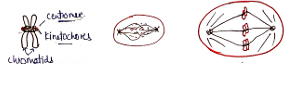
Metaphase: This phase begins with complete disintegration of nuclear envelope.
- Chromosomal condensation completed and two sister chromatids are holds together by centromere.
- Kinetochores (small disc shaped at the surface of centromere). Spindle fibre attach to kinetochores and chromosome are moved to spindle equator and get alligned.
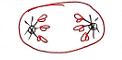
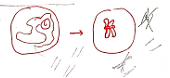
Anaphase: Begins with splitting of each chromosomes. Separation of chromatids and move towards opposite poles.
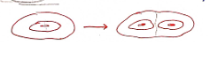
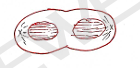
Telophase: Last stage of mitosis and in this, chromosomes loose individuality (decondensation) and chromatin material is collected as a mass at two poles. Nuclear membrane & nucleolus reappear and two nuclear formed. Other organelles reappears.
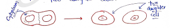
ii) Cytokinesis: It is the division of parent cell cytoplasm after nucleus division to form two daughter cells.
2) Meiosis
It occurs in gamete cells (reproductive cells). In this, offsprings are produced by the fusion of a male gamete and female gamete.
It occurs in two stages:
i) Meiosis - I: It is divided into four phases.
- Prophase-I: Is longer and more complex. It is further sub-divided into five phases:
a. Leptotene phase: Condensation of chromosome starts.
b. Zygotene phase: pairing of chromosomes, and the paired chromosome refered as Homologous chromosomes. This process is known as Synapsis and formed synaptonemal complex. Bivalent/Tetrad formation.
c. Pachytene phase: exchange of genetic material b/w two homologous chromosomes i.e. Crossing over.
d. Diplotene phase: dissolution of synaptonemal complex and formed X-shaped structure termed as chiasmata.
e. Diakinesis phase: Terminalisation of chiasmata, chromosome condensation completed, mitotic spindle is assembled, Nucleolus and nuclear envelop disappear.
Metaphase-I: chromosomes allign at the equatorial plate, microtubules are seen attaching to the pair.
Anaphase-I: Homologous chromosomes separated.
Telophase-I: Nuclear membrane & nucleolus reappear.
This telophase followed by cytokinesis and formed two haploid (gametes) cells.
Interkinesis: It is the stage between two meiotic divisions and usually lasts for a short period of time.
ii) Meiosis - II: It is similar as normal mitosis. It also comprises of four stages:-
- Prophase-II: condensation of chromosomes, formation of spindles and disappearance of nucleus.
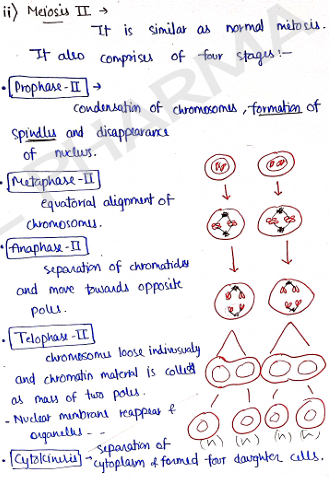
- Metaphase-II: equatorial alignment of chromosomes.
- Anaphase-II: separation of chromatids and move towards opposite poles.
- Telophase-II: chromosomes loose individuality and chromatin material is collected as mass of two poles. Nuclear membrane reappear & organelles.
- Cytokinesis: separation of cytoplasm & formed four daughter cells.
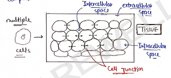
CELL JUNCTION
It is the contact/connection between the neighboring cells or between a cell and the extracellular matrix.
- These are made up with multiprotein complex.
Functions:
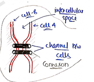
* they creates the communications between the cells.
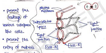
* they maintain the paracellular barrier and prevent the movement of unwanted water solutes and other substances.
* helps in attachement of cells and also responsible for transfer of substances & ions.
* also provide strength to the cells.
Types:
They are divided into five parts:-
- Tight Junction
- Adherens Junction
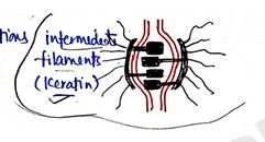
- Desmosome
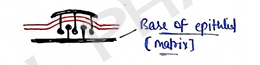
- Hemi-desmosome
- Gap Junction
i) Tight Junction:
ii) Adherens junction:
iii) Desmosomes:
iv) Hemi-desmosome:
v) Gap Junction:
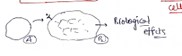
CELL COMMUNICATION
Also known as "Cell Signalling".
- It is the communication b/w the cells. In this, the cell transmit (send), receive and process the signal from one cell to another cell or with itself.
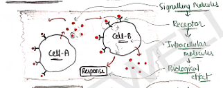
* Cell-A $\to$ Signalling Molecules $\to$ Cell-B (Receptor) $\to$ Biological effect.
- It is important to maintain homeostasis, for growth and development of cells.
- they communicate with each other to help in transport substances, generate electrical potential across cell membrane and respond to change occuring in internal & external environment.
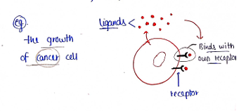
General principle: In cell communication, one cell send it signals (i.e. Ligands) which received by the receptor of another cell, then receptor activates and give response target cell through secondary messengers (eg. cAMP, $IP_3$).
- Reception Transduction Response
Intracellular signalling pathway activation by extracellular signal molecule: Cell communication is of two types:-
- Intercellular signalling (extracellular): It refers to the communication between the cells.
- Intracellular signalling: It refers to the pathway which involves various chain reactions within the cell.
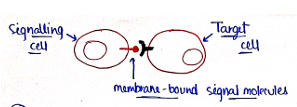
Forms of Intracellular signalling: These are of following types:- a) Contact-dependent b) Paracrine c) Synaptic d) Endocrine e) Autocrine signalling
a) Contact-dependent:
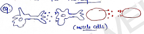
b) Paracrine signalling:
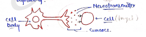
c) Synaptic signalling:
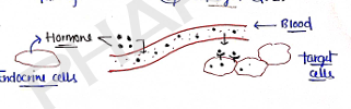
d) Endocrine Signalling:
e) Autocrine signalling:
TISSUE LEVEL OF ORGANISATION
CHAPTER - 3rd
UNIT - 1st
Syllabus: Classification of tissue, structure location and functions of Epithelial, muscular, Nervous and Connective tissue.
Tissue: It is a group of some cells which have similar structure and functions. * each tissue carries out a unique functions in body. * They are only found in multicellular organism.
Histology It is the branch of science that deals with the study of tissue.
Functions:
- All tissue together performs the functions to support the body system.
- Tissue provides shape to the body and help body to store energy.
- Tissue helps in the formation of various organs. (eg Heart, kidney, lungs etc..)
Classification of Tissue Tissues are classified into four major types based on their structure and functions.
TISSUE
- Epithelial tissue - provides covering and protection to the body.
- Connective tissue - provides structural framework to the body.
- Muscular tissue - provides movement to body.
- Nervous tissue - responsible for coordination and communication.

CLASSIFICATION OF TISSUE
(1) EPITHELIAL TISSUE
- SIMPLE TISSUE
- Simple Squamous
- Simple Cuboidal
- Simple Columnar
- Simple Ciliated
- STRATIFIED TISSUE
- Stratified Squamous
- Stratified Cuboidal
- Stratified Columnar
- Transitional
- GLANDULAR TISSUE
- Endocrine
- Exocrine
(2) CONNECTIVE TISSUE
- LOOSE CONNECTIVE T.
- Areolar connective
- Adipose connective
- Reticular connective
- DENSE CONNECTIVE T.
- Dense regular
- Dense irregular
- Elastic
- CARTILAGE
- BONE
- BLOOD
(3) MUSCULAR TISSUE
- SMOOTH
- CARDIAC
- SKELETAL
(4) NERVOUS TISSUE
- NEURONS
- GLIAL CELLS
1. EPITHELIAL TISSUES
Also known as 'Vascular tissue' or 'Epithelium'.
- These are those tissue which are made up of closely packed cells and form continuous sheet.
- They contain minimal extracellular space but they are arranged on basement membrane which is made up by thin sheet of connective tissue.
- They mainly form outer covering of skin/body and internal organs like kidney, lungs, glands etc.
FUNCTIONS:-
- They provide covering to our body and various internal organs.
- They provide protection to our body from mechanical injury, harmful chemicals, loss of water.
- They helps in secretion of various hormones and chemicals through glands.
- They helps in absorption of nutrients from food.
- They also helps in excretion of waste products.
CLASSIFICATION:- They are classified on the basis of their Structure:
- Simple
- Stratified
- Glandular
SIMPLE EPITHELIAL TISSUE
These are those tissue which are made up with single layer of cells.
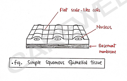
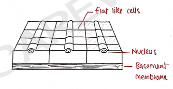
i) Simple Squamous epithelium
- It is made up of only a single layer of flat, scale like cells.
- the nucleus of each cell is oval/spherical.
- Location - Heart, blood vessels, Lymphatic vessels, Airsacs of lungs, Lining of kidney.
- functions - Blood filteration in kidney, Diffusion of to blood vessels, exchanges of gases [$O_2/CO_2$], secreting substances.
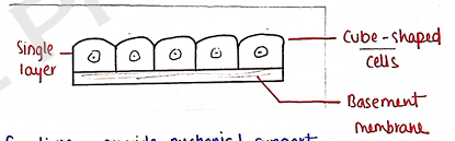
ii) Simple Cuboidal Epithelium
- It is made up of a single layer of cube shape cells, that rest on a basement membrane.
- Location - Surface of ovary lines, kidney tubules, thyroid glands, ducts of many glands.
- functions - provide mechanical support, secretion and excretion.
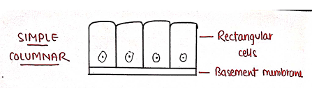
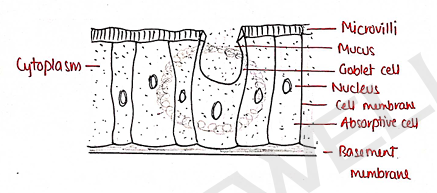
iii) Simple columnar Epithelium
- It is made up of single layer of rectangular cells arranged on basement membrane.
- It contain goblet cells, cilia and microvilli.
- Location - lining of stomach, intestine, uterus, uterine tubes and some parts of respiratory tract.
- functions - secretion and absorption, excretion and protection.
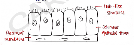
iv) Simple Ciliated Epithelium
- Also known as pseudostratified columnar epithelium. It is made up of only a single layer and have irregularly shaped columnar cells.
- Location - few portion of upper respiratory tract, ventricles of the brain, spinal cord.
- functions - protection and secretion, moves mucus and other substances by ciliary action.
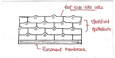
2. STRATIFIED EPITHELIAL TISSUE
These are those tissue in which cells are arranged in multiple layers. i.e. one over another.
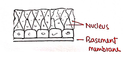
i) Stratified Squamous Epithelium
- It is made up of multiple layers of flattened squamous cells.
- Location - skin, oesophagus, pharynx, lining of mouth, tongue, vagina
- It is of two types:
- Keratinised - contain keratin fibres, provides waterproof, protective qualities to the skin.
- Non-keratinised - it remain moist, such as vagina, mouth and oesophagus.
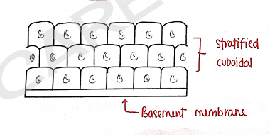
ii) Stratified Cuboidal Epithelium
- It is made up of two or more layers of cube shaped cells.
- Location - Sweat gland, Salivary glands and mammary glands.
- functions - Protection, Secretion of saliva, sweat, and milk.
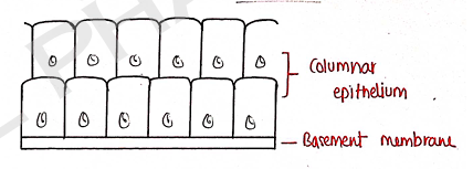
iii) Stratified Columnar Epithelium
- It is made up of multiple layer of rectangular shaped cells.
- Location - Urethra, Oesophageal gland, mucus membrane, Lining of eyelids.
- functions - Protection and secretion.
iv) Transitional Epithelium
- It is presented at the sites which are subjected to changes in stress and tension.
- Location - urinary bladders, their lining.
- functions - provide contraction. (Walls of Urinary bladders.)
3. GLANDULAR EPITHELIUM
These are those tissue which are used for their secretion action, in which they excrete various hormones and chemicals.
- They secreted into ducts, surface of the body, or directly into the blood.
i) Exocrine glands
ii) Endocrine glands
2. CONNECTIVE TISSUE
- These are those tissue which connects or bind different organs or different parts of an organs.
- It is the most diverse and widespread tissue in the human body, found in almost every organ of the body.
- It is arises from the mesoderm layer of embryonic [stem cell tissue].
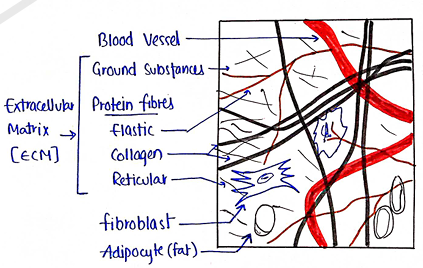
- Connective tissue is composed of large amount of extracellular matrix (ECM), limited number of cells, fluids and number of fibres.
- These all collectively known as ground substances.
- They comprises of cells i.e. plasma cells, WBC, mast cells, adipocytes (fat cells), Macrophages, fibroblast mostly.
- Contain fibres:
- Collagenous fibres are tough and strong (strength).
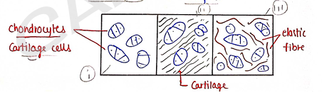
- Elastic fibres are elastic and extensible in nature Branches (elasticity).
- Reticular fibres are delicate / fragile in nature, provide support.
- Connective tissue present in the form of soft, gel-like to firm, flexible and hard type.
FUNCTIONS:
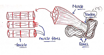
- It connects different tissue of body. (eg muscles are connected with bones by tendons).
- It supports various tissue, organs and structures of the body.
- Blood helps in transportation of and nutrients, and also provide defence system for body.
- It work as structural framework of body.
TYPES OF CONNECTIVE TISSUE:
- Loose C.T. (Areolar, Adipose, Reticular)
- Dense C.T. (Regular, Irregular, Elastic)
- Others (Cartilage, Bones, Blood)
1. Loose connective tissue :-
These are those tissue in which cells are loosly arranged with fibres or ground substances in Matrix. It is well vascularized and provides blood supply to nearby epithelial tissue. It is one of the most widely distributed tissue which connects several body structure by acting as elastic glue which allow movement.
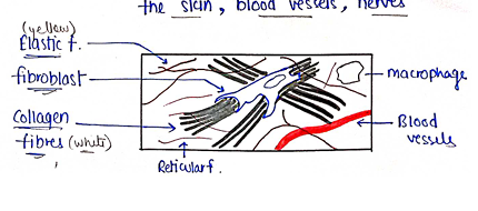
- Areolar Connective Tissue: (strength, connectivity)
- It connects the skin to the underlying structures. It works same as loose connective.
- Location - found between muscles, below the skin, blood vessels, nerves.
- Adipose Connective tissue:
- It contain adipocytes (fat cells), which store energy (glucose) for the body.
- Location - under the skin, b/w internal organs, bone marrow.
- Structure -
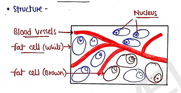
* **functions** - store energy in the form of fat, support and protection. Brown fat produce heat generation.
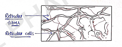
- Reticular tissue:
- The term reticular means 'like a net'. The reticular network is formed by thin, branches of reticular fibres.
- Location - found in spleen, lymph nodes, and bone marrow.
- functions - provide protection and helps in the production of blood cells.
2. Dense connective tissue / Dense fibrous :
These are those tissue which are densely packed and form rope like structure. They mainly contain fibrocytes, fewer fibroblast cells, and fibres in large amounts.
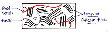
- Dense Irregular Tissue:
- In this, collagen fibres are arranged in irregularly (random).
- It is arranged in the form of thick mat like strong connective tissue.
- (Eg) Dermis layer of skin, outer covering of organs such as kidney, spleen etc..
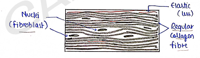
- Dense regular tissue:
- In this, collagen fibres are arranged in parallel, regular.
- they provide strong attachment, flexibility.
- (Eg) It is present in tendons (attach muscles to bone) and ligaments (attach bone to bone).
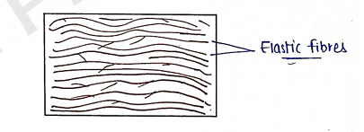
- Elastic Dense tissue:
- In this, elastic fibres are arranged parallel to each other.
- It provides elasticity with strength.
- (eq) Trachea, Bronchi, lungs and ligaments. Arteriolat walls are also made up of elastic fibrous tissue.
3. Cartilage :-
It is strong, flexible connective tissue. It contain only one type of cells i.e. chondrocyte which produces the fibres and the tough, rubbery ground substance of cartilage.
- It protects joints and bones.
- It is present at the end of bones and helps in the formation of bones.
- It is of three types:-
- i) Hyaline Cartilage tissue - It forms the covering of ends of bones and it founds in rings of trachea.
- ii) Fibro cartilage tissue - The Strongest and most durable tissue, It forms intervertebral disc of vertebral column. It also present in knee joints and work as shock absorber.
- iii) Elastic Cartilage tissue - It contain less amount of collagen fibre and large elastic fibres, which provides flexibility. It present in external ear and larynx.
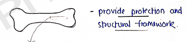
4. Bone (Osseous tissue):
It is the hard connective tissue, that contain a high concentration of salts like Calcium phosphate and Calcium Carbonate (minerals). It also consists of collagen fibres. It provide protection and structural framework.
- It presents in Arms, legs, Ribs etc..
5. Blood:
It is liquid connective tissue which are formed in the bone marrow and other tissue.
- It is composed of 55% plasma and 45% cells.
- Blood cells are RBCs (Red blood cells), WBCs (White blood cells), Platelets.
- Helps in connect different tissue.
- helps in transportation of gases, nutrients, drugs etc..
- Body defence System.
[Diagram: Blood cells, platelets]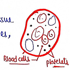
3. MUSCULAR TISSUE
These are those tissue which is made up from muscle fibres and helps in the movement of body.
The main functions of this tissue is Contraction and relaxation, which helps in movement.
FUNCTIONS:
- They allow movement of bones and joints through contraction & relaxation.
- Helps in the production of heat (skeletal muscle).
- It maintain body posture.
- It forms protective layer around organs.
- It play a major role in pumping of blood by the heart, peristaltic movement of stomach, movement of food in GIT etc.
- It helps to express feeling.
TYPES OF MUSCULAR TISSUE: Based on the location, muscular tissue divided into three parts :-
- Skeletal muscles
- Smooth Muscles
- Cardiac Muscles
i) Skeletal Muscles:
- These are those muscles which are attached to the bones and helps in the movement of bones and joints.
- These are cylindrical shaped, multinucleated cells having a group of muscle fibrils.
- Also known as striated as they contain strips.
- These are voluntary in nature which is controlled by Somatic Nervous system.
- present upon the skeletal system.
- It comprises 40% of body masses.
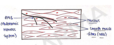
ii) Smooth Muscles:
- These muscles are thin and spindle shape. They consists of actin (thin) and myosin (thick) filaments sliding over each other and provide contraction.
- they are unstriated muscle fibres, having a single nuclei.
- Involuntary in nature and are controlled by Autonomic Nervous system.
- Location Iris of the eyes, Blood vessels, lungs, stomach, gall bladder, Intestine.
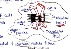
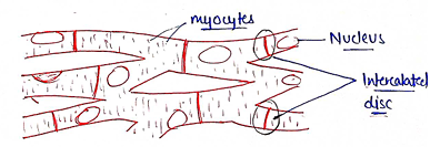
iii) Cardiac Muscles:
- These are those muscles which are found in heart (myocardium).
- It consist of branched striated fibres with one or two centrally located nuclei.
- They have involuntary control which have automatic and rhythmic contraction of muscles.
- functions:- pump blood to all part of the body, helps in generating contraction, work as protective layer for heart.
4. NERVOUS TISSUE
These are those which are found in the brain, spinal cord and nerves and are responsible for coordinating and controlling many body activities.
The main function of nervous tissue is to receive information from stimuli, analyze with brain/spinal cord and send response.
TYPES: It consists of two types of cells
- Neurons/Nerve cells.
- Glial Cells/Neuroglia cells.
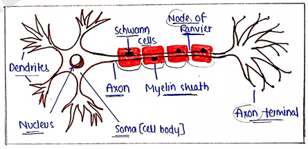
1. Neurons/Nerve Cells: It is the structural and functional unit of Nervous tissue. It is responsible for all the functions provided by Nervous tissue.
FUNCTIONS:
- Responsible for coordination and communications.
- Regulate and controls body functions.
- Send and receive impulses (information).
- Stimulates muscle contraction.
- play major role in emotions & memory.
It contain various parts:
- Cell body (Soma) - main body contain nucleus & dendrites.
- Dendrites - Branches, receive signals & passed.
- Axon - passes signal.
- Myelin sheath - jump the message/fast.
- Axon terminal - end part, transmit signal.
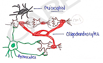
2. Neuroglial/Glial Cells: These are supporting cells which provides connects, support and regulate the functioning of neurons.
- It is of three types -
- Astrocytes - regulates the functions and protection.
- Microglia - destroy pathogens.
- Oligodendrocytes - enhance conduction speed.
Chapter 1
Chapter 2
Chapter 3